03 Proteins I An Introduction to Protein Structure and Function
Outline
3.1 Amino acid chemistry
3.2 Proteins are polymers of amino acids
3.3 Proteins are molecules of defined shape and structure
3.4 Examples of protein structures and functions
Learning Objectives
- Compare the 20 amino acids commonly found in proteins with each other in terms of their chemical properties.
- Describe the general properties of proteins and peptides.
- Analyze the different levels of protein structure.
- Describe several examples that illustrate the diversity of protein structures and functions.
Section 3.1 Learning Objective
Compare the 20 amino acids commonly found in proteins with each other in terms of their chemical properties.
Amino Acids
The building blocks of proteins
Contain amine and carboxylic acid functional groups
There are 20 amino acids, all with different side chains (residues).
Figure 3.2 Structure of an amino acid.
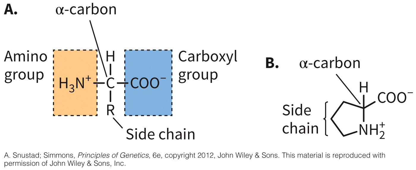
Alpha carbon and the side chain (R group)
There are 19 alpha amino acids (amino group, a central alpha carbon, and a carboxyl group) and 1 imino acid (proline – contains a secondary amine)
Amino Acids
The majority are achiral.
Figure 3.3 Chirality of amino acids.
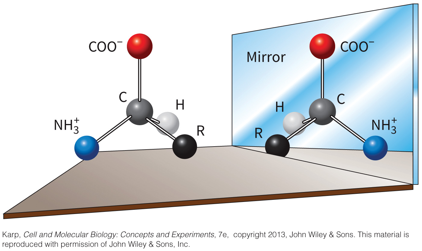
NOTE: Nature prefers the L-enantiomer for amino acids
Zwitterion Defined
Zwitterion is a molecule that possesses both a positive and negative charge.
- An amino acid at neutral pH is an example.
Figure 3.4 Zwitterionic nature of amino acids.
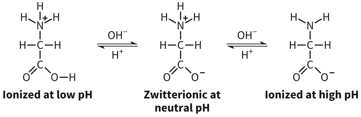
Amino acids can have different charges at different pHs
Amino Acid Classification
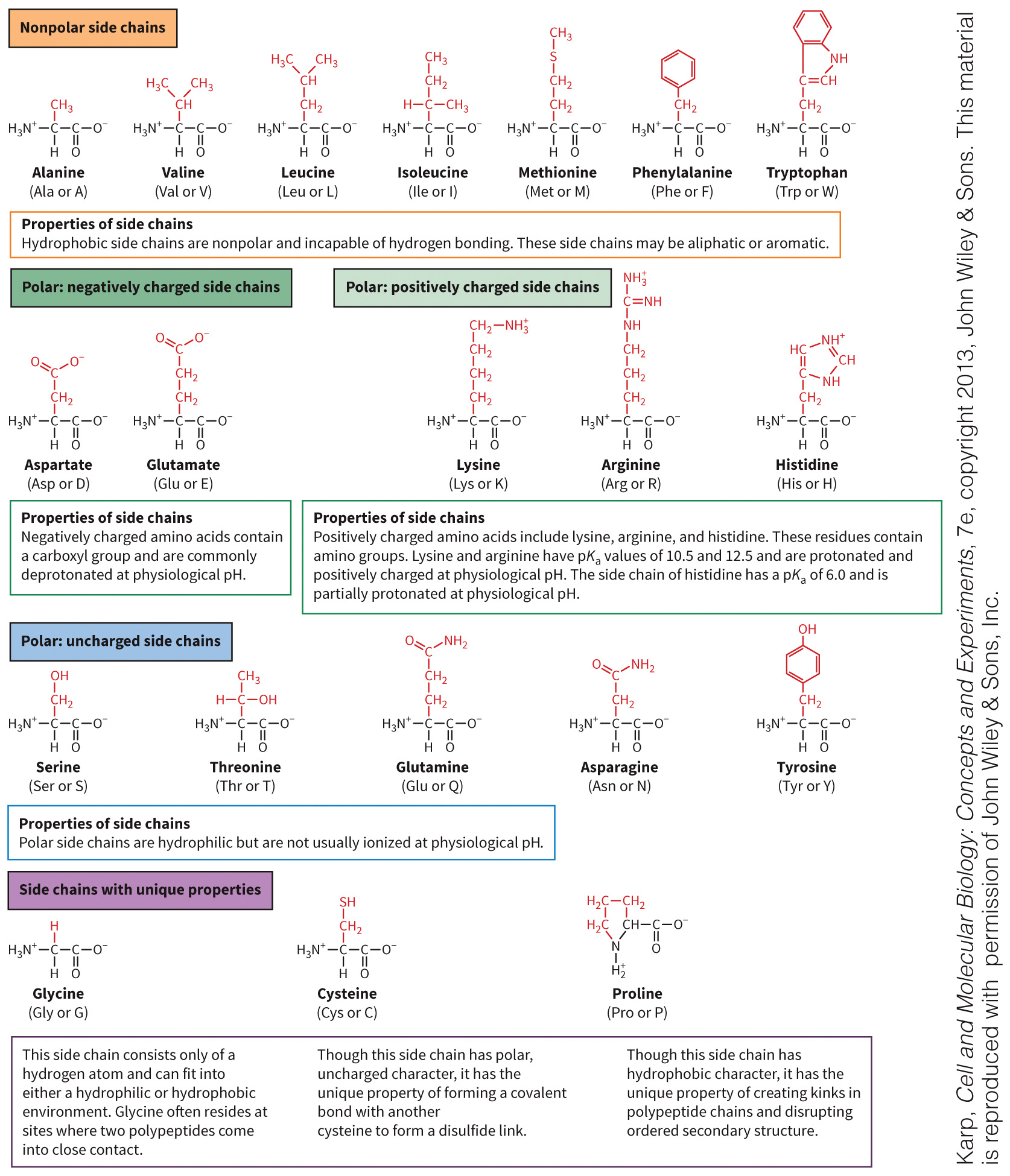
Nonpolar (hydrophobic)—usually an alkyl group, an aromatic ring, hydrogen, or a nonpolar collection of atoms.
Polar—the side chain is polar or ionic.
- acidic—the side chain is a carboxylate ion.
- basic—the side chain is an amine.
- neutral—the side chain in an alcohol, thiol, or amide.
Branched chain amino acids (BCAAs) = Valine, leucine and isoleucine.
Polar acidic amino acids have negatively charged side sides, whereas lysine, arginine, and histidine have positively charged side chains
Amino Acid Structures
Figure 3.5 Amino acids.
Side Chain Ionization
Amino acid side chains play a pivotal role in protein folding and protein–protein interaction.
These side chains may become ionized at different pKa values.
This has an effect on overall protein structure.
The pKa Values of Amino Acid Side Chains
| Amino acid | Three-letter abbreviation | One-letter abbreviation | pKa |
|---|---|---|---|
| Amino | - | - | 9.30 |
| Arginine | Arg | R | 12.48 |
| Lysine | Lys | K | 10.53 |
| Tyrosine | Tyr | Y | 10.07 |
| Cysteine | Cys | C | 8.18 |
| Histidine | His | H | 6.00 |
| Glutamate | Glu | E | 4.25 |
| Aspartate | Asp | D | 3.65 |
| Carboxyl | - | - | 2.10 |
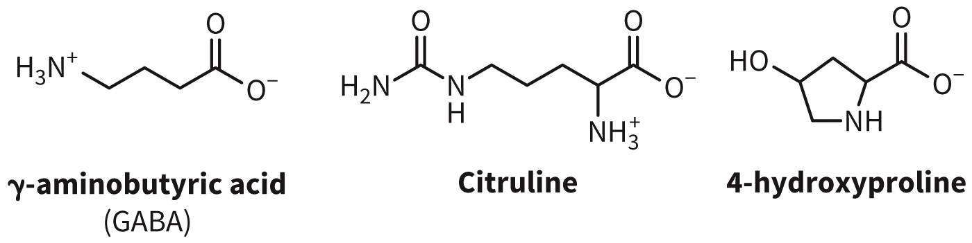
Roles of Amino Acids in Biochemistry
Amino acids serve a variety of roles in biochemistry including:
- lipid metabolism
- amino acid catabolism
- neurotransmission
Figure 3.6 Amino acids not commonly found in proteins.
Section 3.2 Learning Objective
Describe the general properties of proteins and peptides.
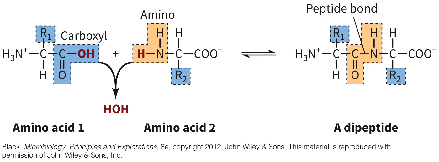
Peptide Bond Defined
Peptide bond is the linage between two amino acids.
- This is an amide linkage between the carboxyl group of amino acid and the amino group of the next.
- A water molecule is removed in order for an amide bond to form.
Figure 3.7 Peptide bond.
Typically in a trans conformation to prevent steric hindrance
Peptides Defined
Peptides are linear polymers of amino acids.
- Dipeptides have two linked amino acids.
- Oligopeptides have 4–20 amino acids.
- Polypeptides contain 20 or more peptides linked together.
- Proteins tend to be greater than 100 amino acids.
Monomers versus Multimers
Monomers
- Single chains of amino acids folded into an active protein
Multimers - Consist of several subunits
- Subunits labeled based on function (i.e., catalytic and regulatory)
Cofactors Defined
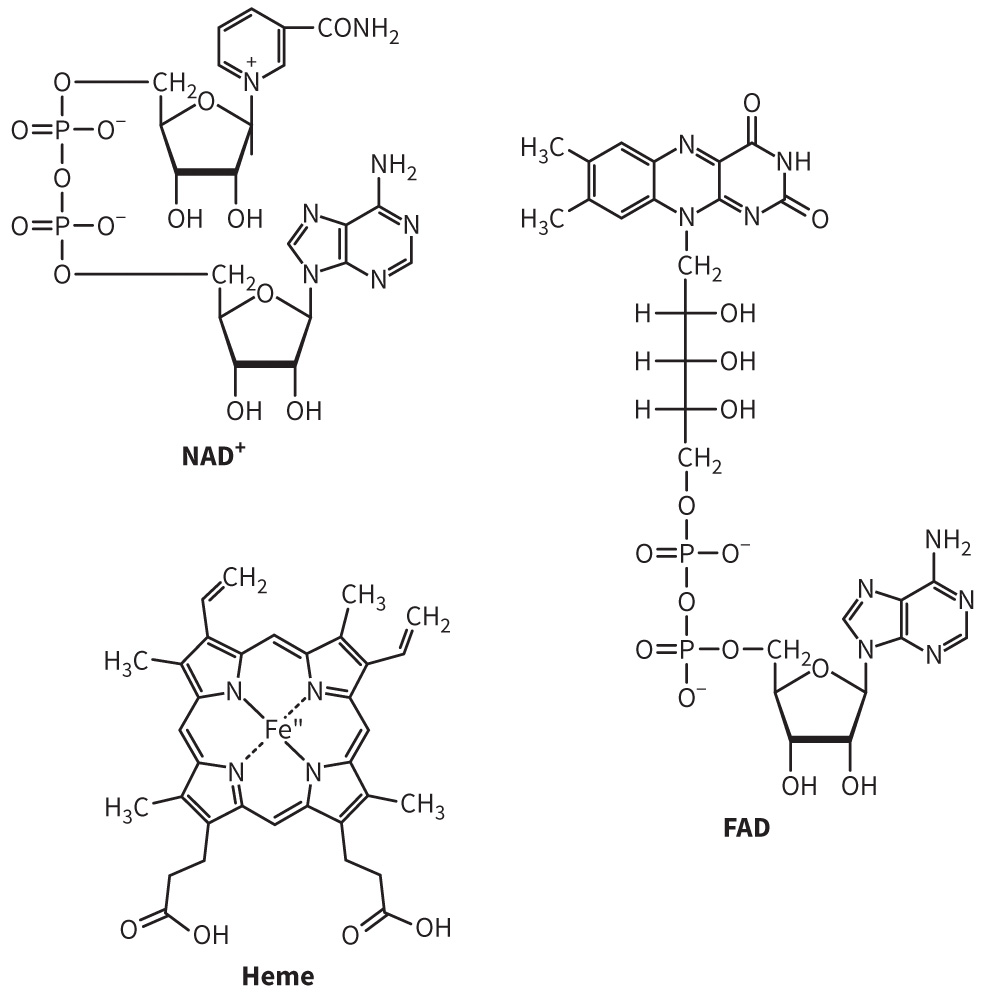
Cofactors are nonprotein groups required for protein binding or activity.
- Can be divided into two categories: minerals (metal ions) and vitamins (small organic groups)
- Metal ions (Ca2+, Mg2+, Zn2+, Fe2+, and Cu2+)
- Organic groups (NAD+ and FAD, pyridoxal phosphate)
- The difference between apoproteins ( do not contain cofactor) and holoproteins (complete protein)
Examples of Cofactors
Figure 3.12 Examples of cofactors.
Amino Acid Modifications
Figure 3.13 Amino acid modifications.
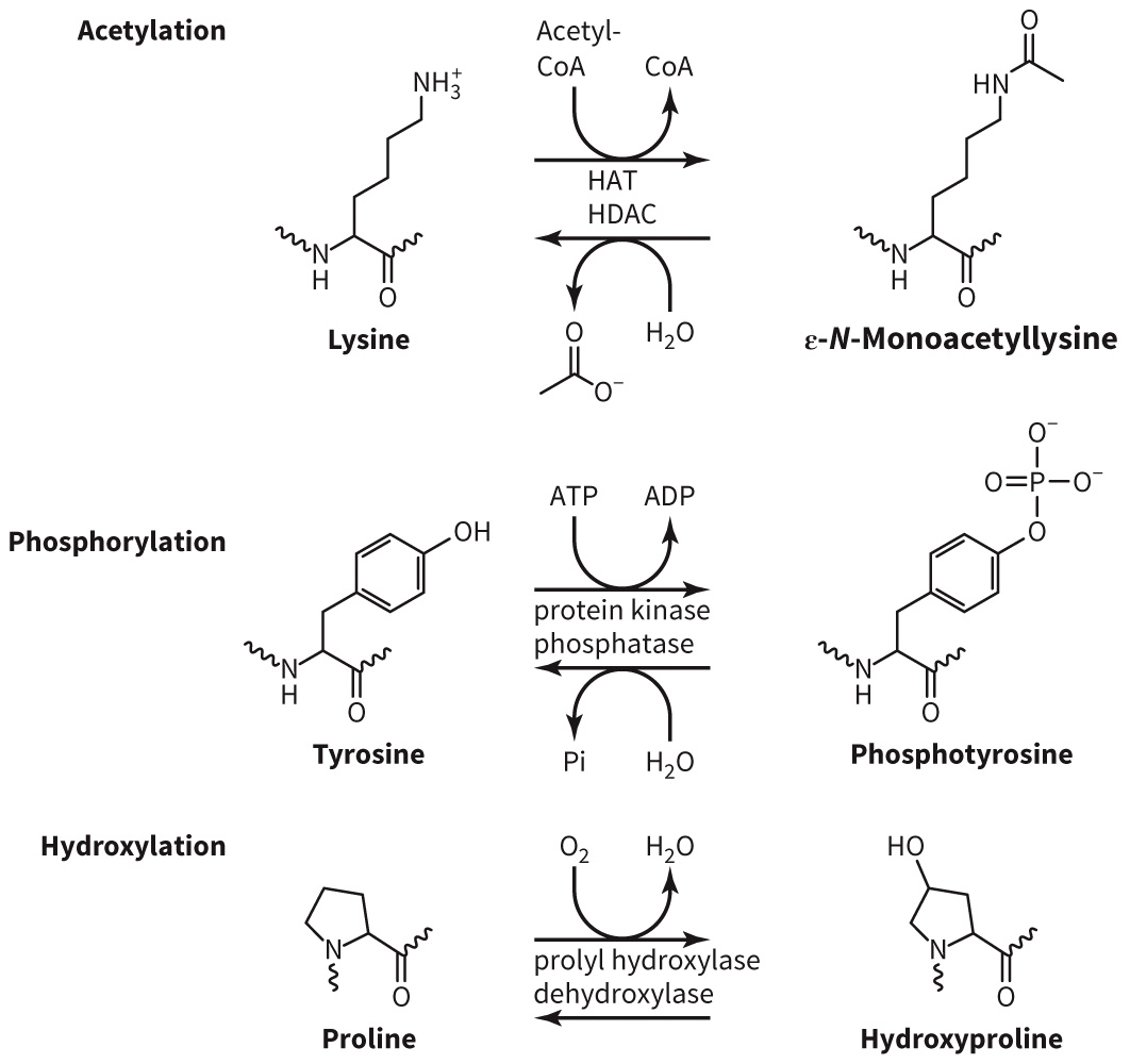
Explain the difference between reversible (phosphorylation) and irreversible (ubiquitination) modifications.
Kinases Defined
Kinases are enzymes that catalyze phosphorylation reactions.
- ATP is typically the source of phosphate.
- Phosphates can add on to the hydroxyl groups of serine, threonine, or tyrosine.
- Phosphorylation can change the activity of an enzyme and is thought to act as a switch in the cell.
Section 3.3 Learning Objective
Analyze the different levels of protein structure.
Four Levels of Protein Structure
Primary
Secondary
Tertiary
Quaternary
Primary Structure
Linear arrangement of amino acids
The order of amino acids is crucial to its function.
Figure 3.14 Primary sequence of a protein.
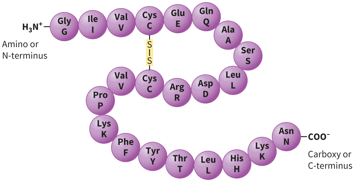
Discuss how substitutions may affect the protein
Conservative substitution = substitutions of the same polarity may not affect function
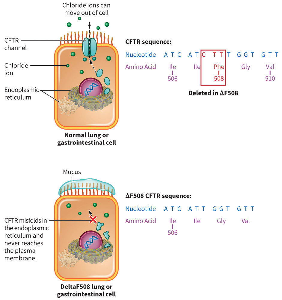
Cystic Fibrosis: An Example of Mutation
Figure 3.16 Cystic fibrosis.
Secondary Structure
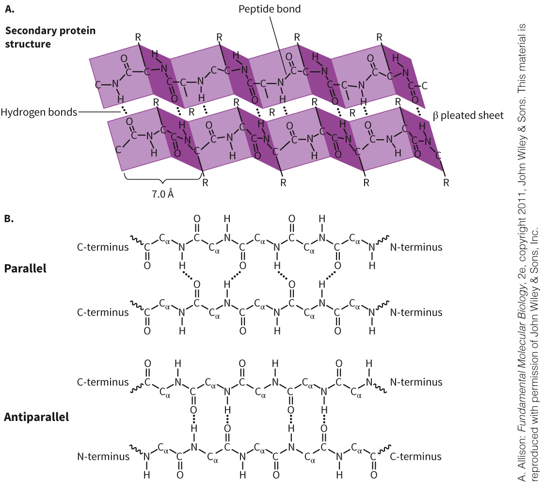
Held together by hydrogen bonding
Consists of two major structural elements:
- α helix
- β sheet
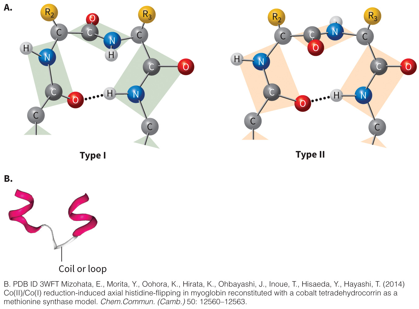
Can also have turns and coils (loops)
Alpha Helix
Most common structure observed in proteins
Figure 3.17A Alpha helix.
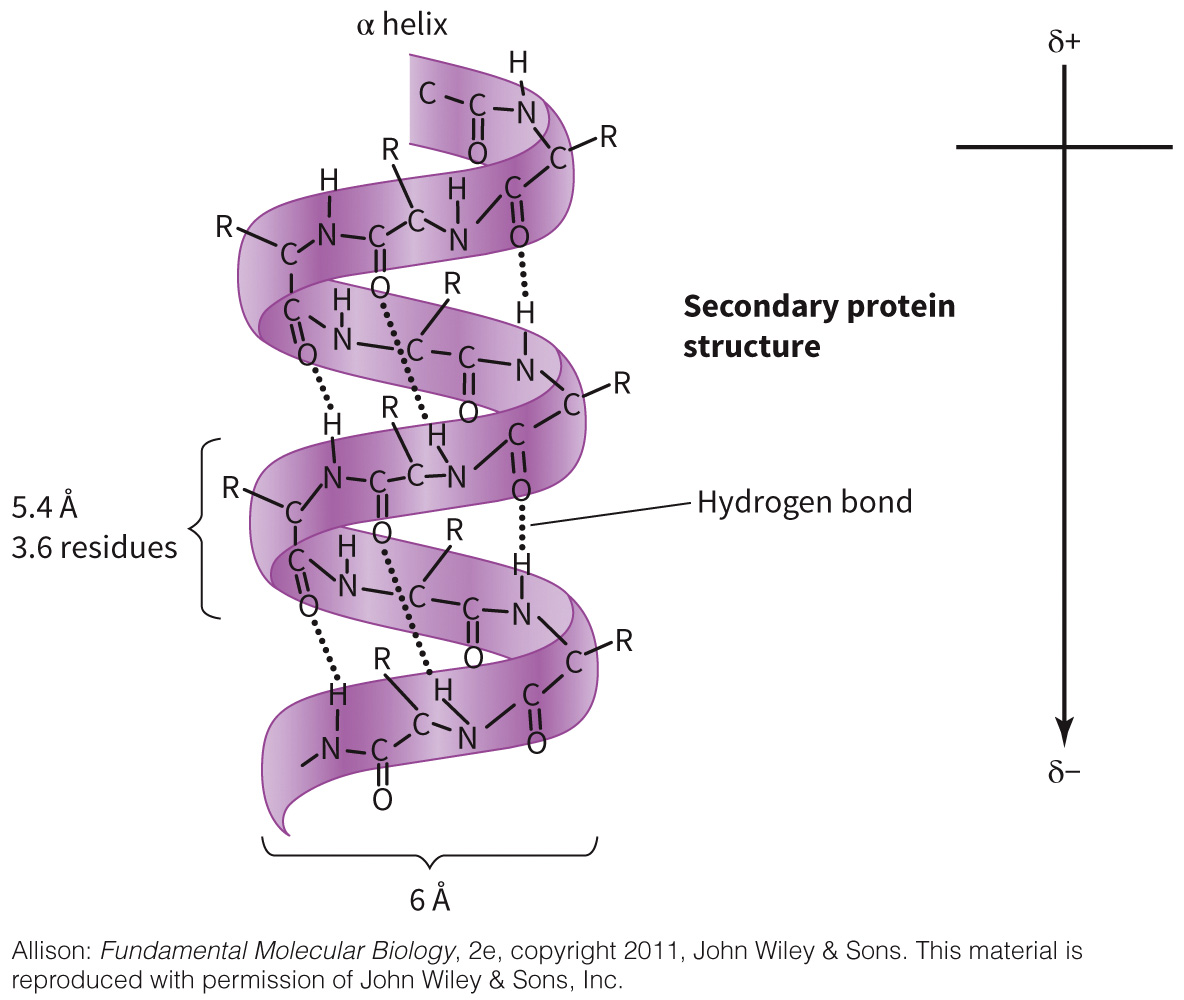
Gly and Pro discourage helix formation
Beta Sheet
Can be parallel or antiparallel
Figure 3.18 Beta sheet.
Hairpin Loops and Turns
Figure 3.19 Turns and coils.
Tertiary Structure
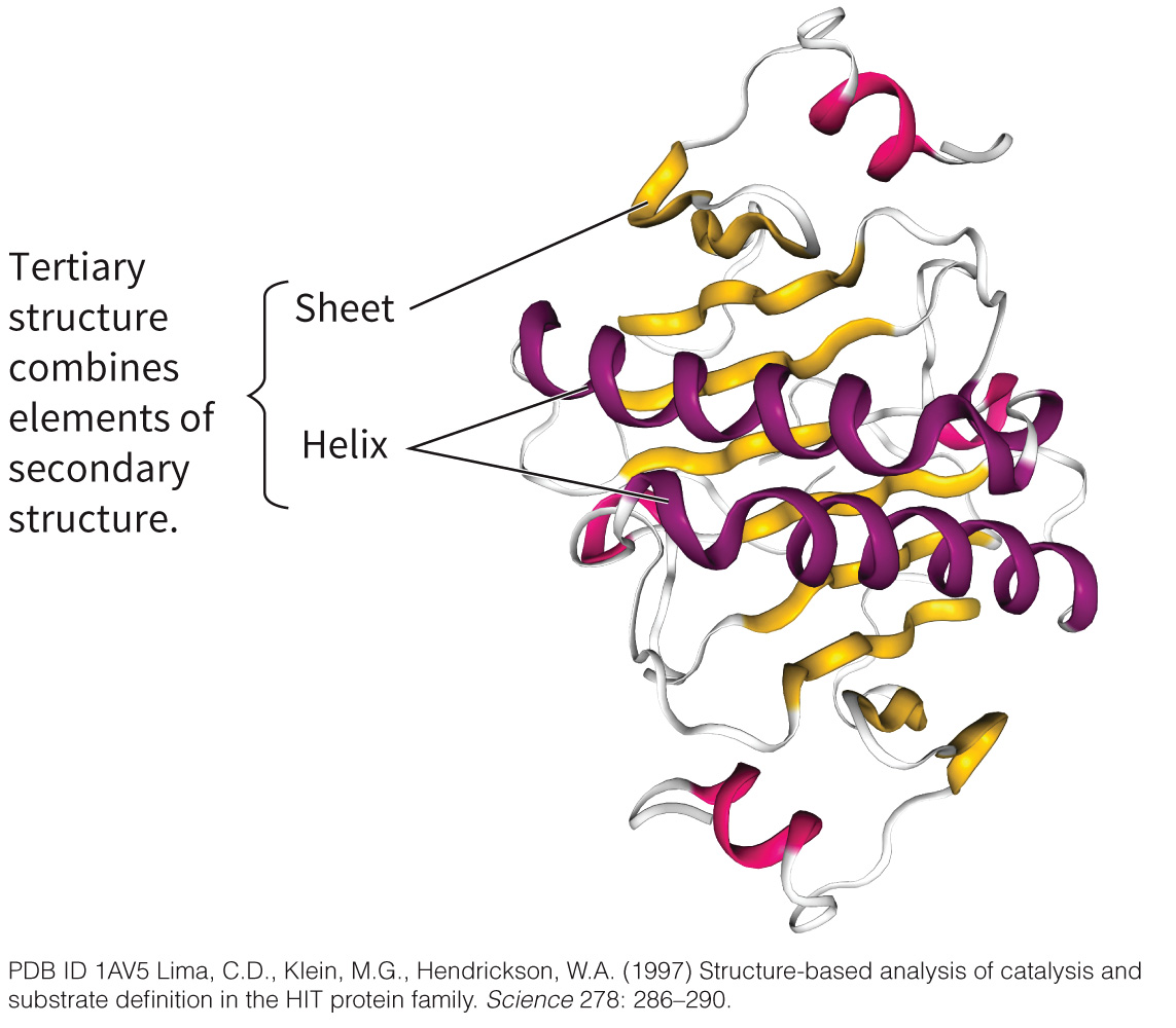
Gives the overall shape of the protein
Figure 3.20 Tertiary structures.
Motifs versus Domains
Motifs
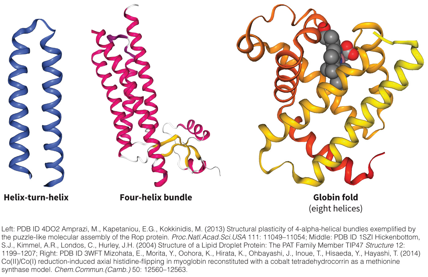
- Combinations of secondary structures
Domains - Pieces of a protein that retain their structure in the absence of the rest of the protein
- A discretely folding structure
Alpha Structures
Figure 3.21 Alpha structures.
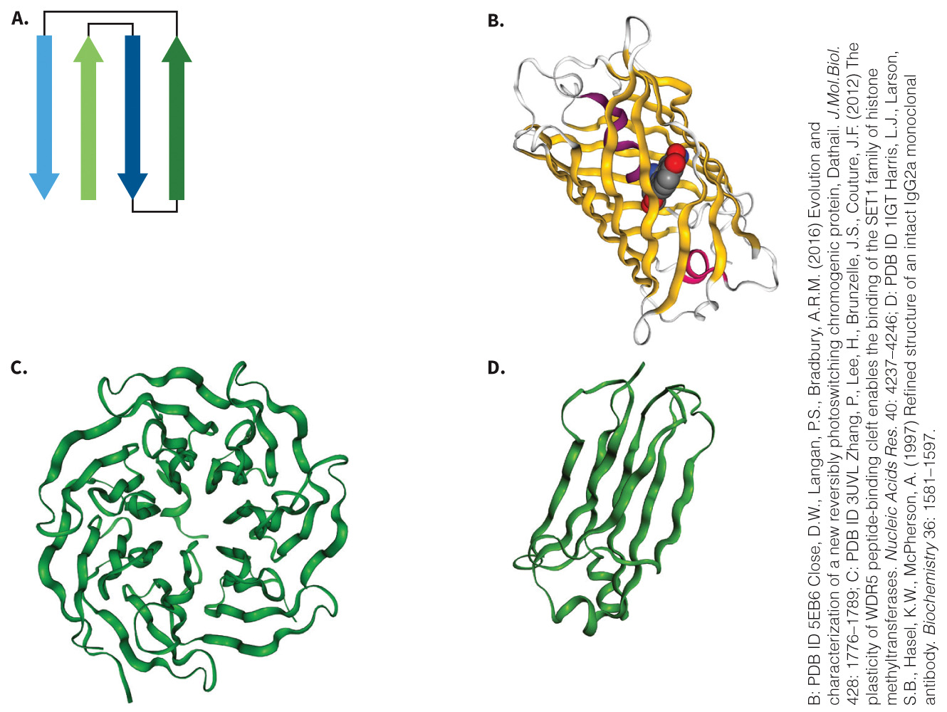
Beta Motifs
Contain mostly β structures
- Include the β barrel, Greek key motif, and β propeller
Figure 3.22 Beta structures.
Bonding Forces Involved in Tertiary Structure
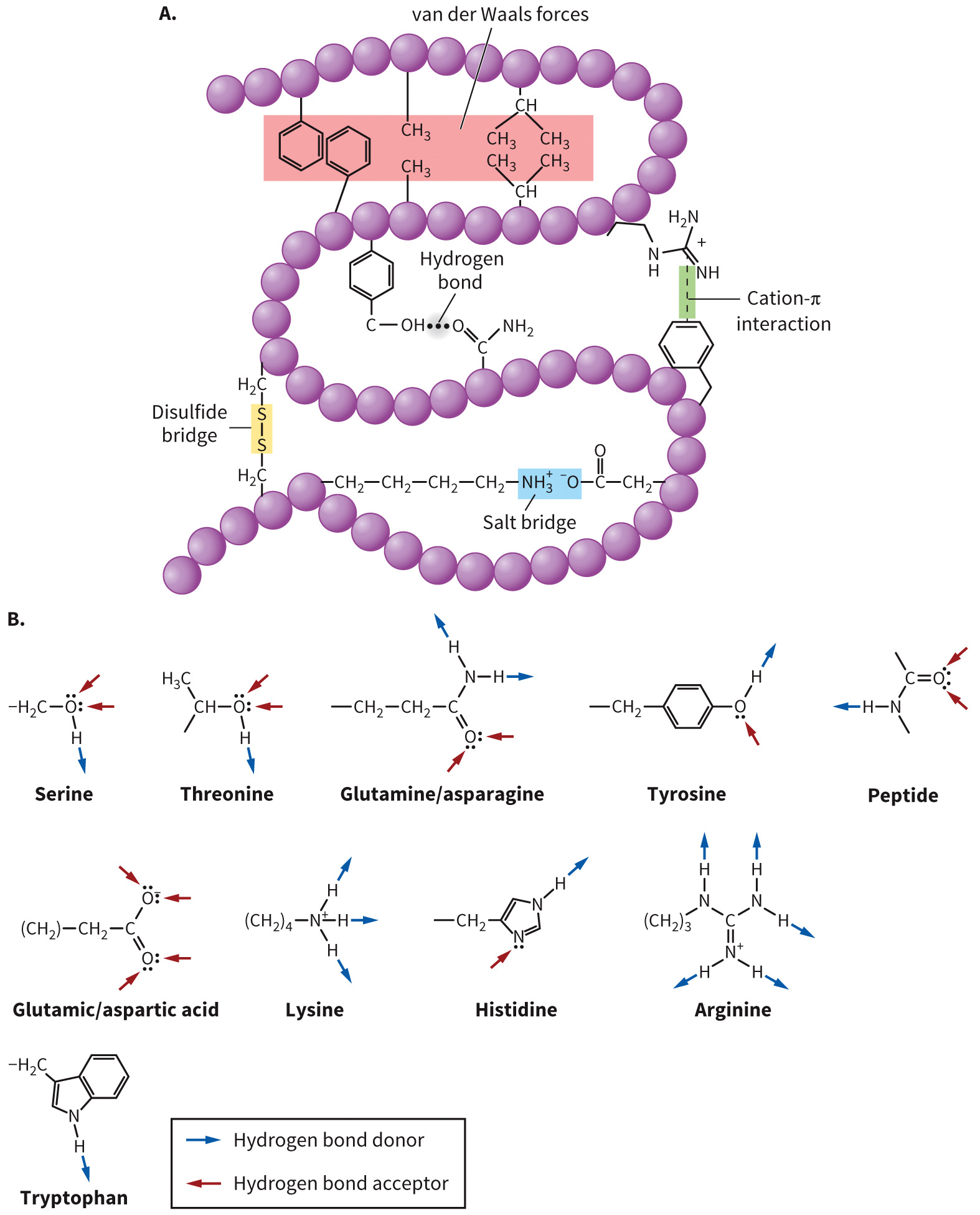
Forces include
- hydrogen bonding
- London dispersion forces
- dipole–dipole interactions
- salt bridges
- cation–π interactions
- disulfide bonds
Explain each type of bonding and point out strongest and weakest forces
Stabilizing Forces Involved in Protein Structure
Figure 3.23 Forces involved in stabilizing proteins.
Hydrophobic Effect Defined
Hydrophobic effect describes the phenomenon in which hydrophobic groups cluster together.
London dispersion and Van Der Waals forces
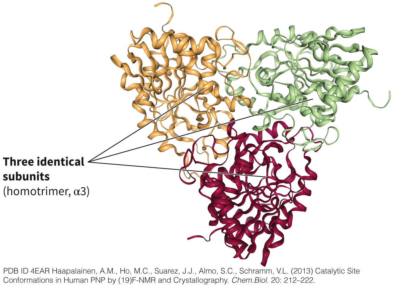
Quaternary Structure
Complex structure that incorporates multiple subunits or different proteins
Figure 3.24 Quaternary structure.
Section 3.4 Learning Objective
Describe several examples that illustrate the diversity of protein structures and functions.
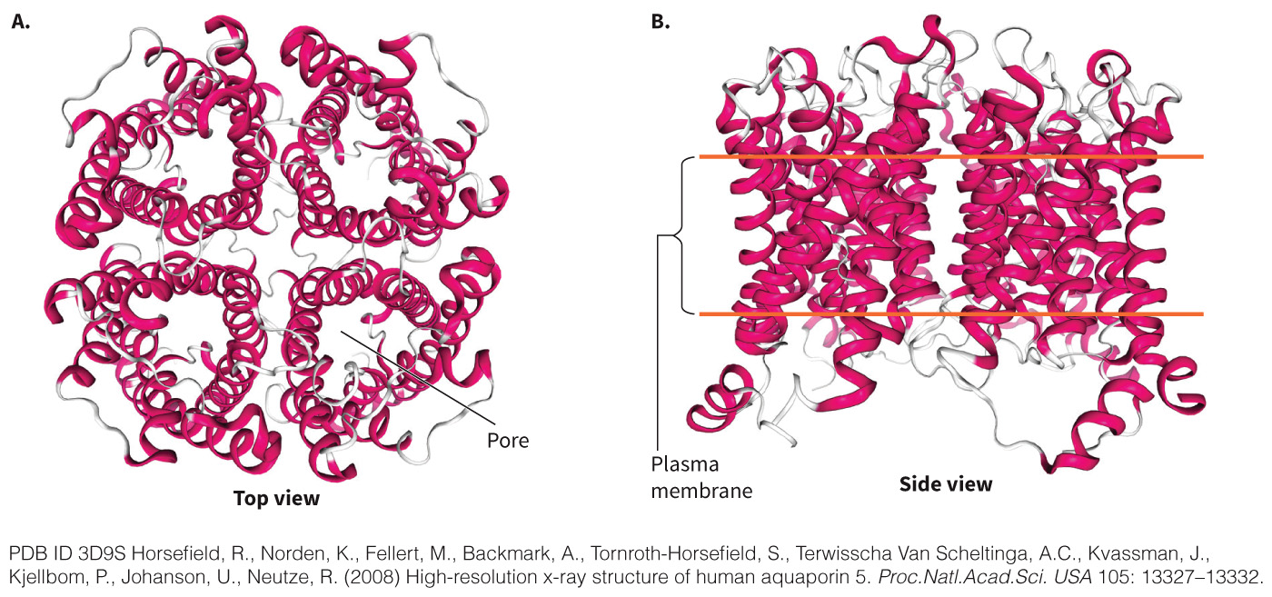
Aquaporin: A Transmembrane Protein
A protein that acts as a pore in the membrane
Selectively allows water to pass in and out of the cell
Consists of six transmembrane α helices
Aquaporin
Figure 3.25 Aquaporin.
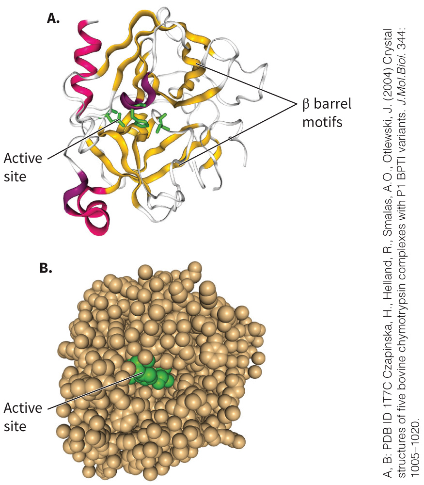
Chymotrypsin: An Enzyme
Produced by the pancreas and secreted into the intestinal lumen
Cleaves dietary protein into peptides
Composed of two β barrels and one short α helix
As an enzyme, contains an active site and regulatory site
Structure of Chymotrypsin
Figure 3.26 Chymotrypsin.
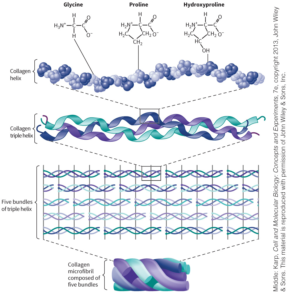
Collagen: A Structural Protein
A fibrous protein mostly concentrated in muscle and connective tissue
Composed of a “triple helix”
Contains glycine (33%), proline (16%), and hydroxyproline (16%)
Also called a collagen helix
Structure of Collagen
Figure 3.27 Collagen.
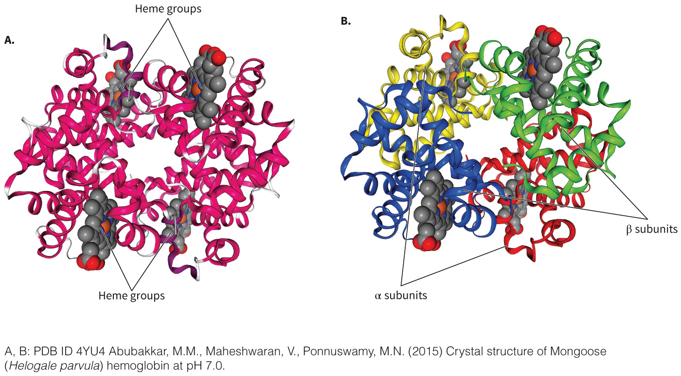
Hemoglobin: A Transport Protein
A globular, tetrameric protein found in red blood cells
Contains four subunits
Transports oxygen to the blood
Uses heme as a cofactor for each subunit
Composed of mostly α helices joined by turns
Hemoglobin Structure
Figure 3.28 Hemoglobin.
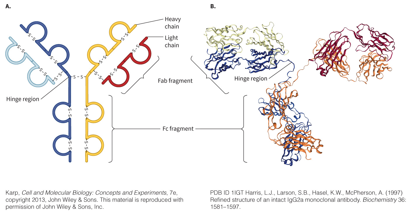
Immunoglobulins: Binding Proteins
Molecules of the immune system that recognize and bind to antigens
Act as adapter molecules between the foreign molecule (antigen) and the immune system cells required to neutralize it
Basic Structure of Immunoglobulins
Figure 3.29 Immunoglobulins.
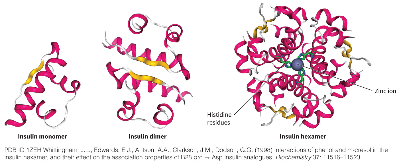
Insulin: A Signaling Protein
A protein hormone produced by pancreatic β cells
A growth factor that signals cells to store energy and divert energy to growth
Interrupted or impaired signaling can result in diabetes
Discuss the difference between Type I (lack of insulin signal) and Type II (defect in insulin receptor) diabetes
Structure of Insulin
Figure 3.30 Insulin.
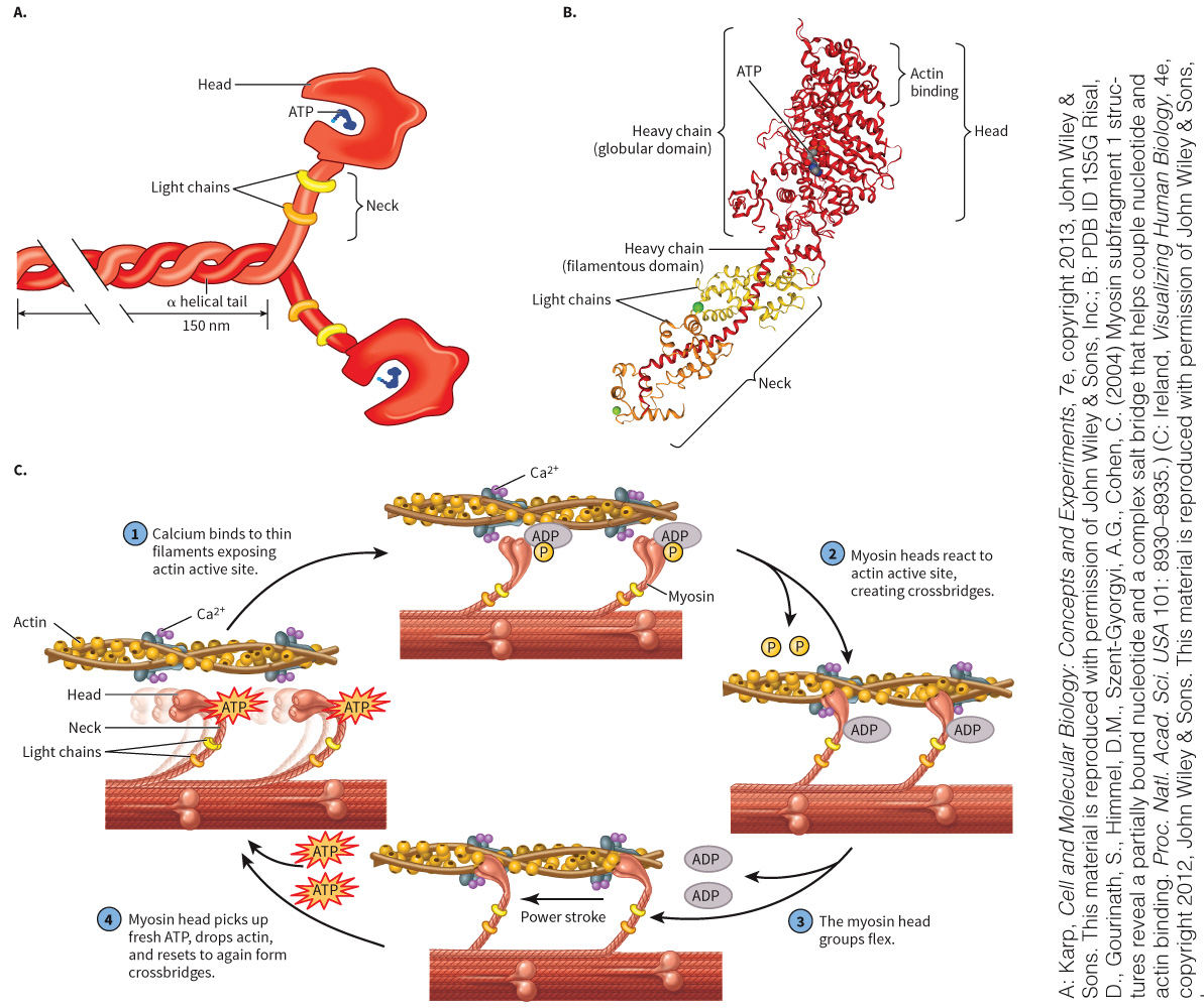
Myosin: A Molecular Motor Protein
Acts as a molecular motor in which energy is used to elicit a muscle contraction and transport vesicles in the cell
Contain heavy and light chains
- Heavy chain—interacts with the cytoskeletal protein, actin
- Light chain—plays a role in binding Ca2+
Mechanism for myosin and movement with ATP
Myosin Structure
Figure 3.31 Myosin.