08 Carbohydrates II Glycogen Metabolism, the Pentose Phosphate Pathway, Glycoconjugates, and Extracellular Matrices
Outline
8.1 Glycogen metabolism
8.2 The pentose phosphate pathway
8.3 Carbohydrates in glycoconjugates
8.4 Extracellular matrices and biofilms
Learning Objectives
- Analyze the pathways of glycogen biosynthesis and breakdown, and the regulation of these pathways.
- Describe the reactions of the pentose phosphate pathway.
- Compare different types of glycosolyated molecules (glycoproteins, glycolipids proteoglycans and peptidoglycans) with regard to structure and function.
- Analyze how extracellular matrices and biofilms are synthesized and function.
Section 8.1 Learning Objective
Analyze the pathways of glycogen biosynthesis and breakdown and the regulation of these pathways.
Glycogen
A stored form of glucose
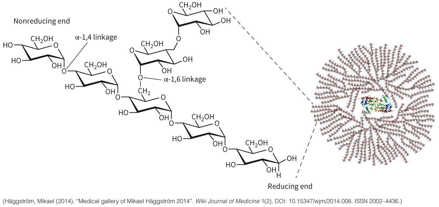
Highly branched polymer of α-1,4 and α-1,6 linked glucose monomers, with a single molecule of the protein glycogenin at the core
It is synthesized and degraded in situ
Storage is regulated by epinephrine and norepinephrine
Acts as a buffer by storing glucose when glucose is plentiful and releasing it at other times when the body needs it
Skeletal muscle stores 75% of the body’s glycogen, the liver contains the rest
Structure of Glycogen
Figure 8.2 Glycogen is a storage form of glucose.
Glycogenesis Defined
Glycogenesis is the synthesis of glycogen.
Requires several important components
- Glycogenin (core protein)
- Glycogen synthase
- Branching enzyme
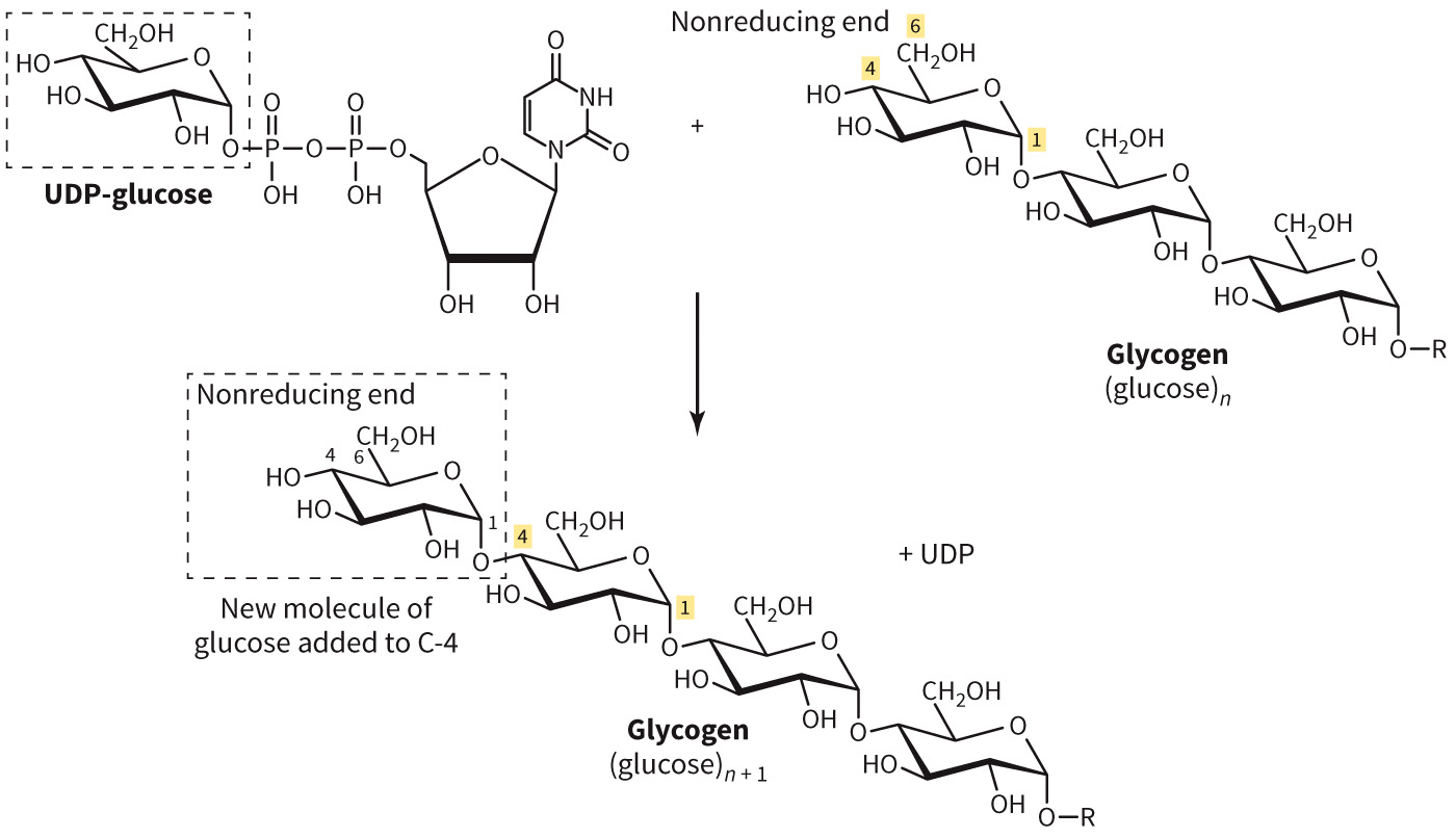
Glycogenin is glycosyl transferase that adds glucose molecules (UDP glucose) to the active site tyrosine – extension occurs until 4 – 13 glucose monomers long then glycogen synthase takes over
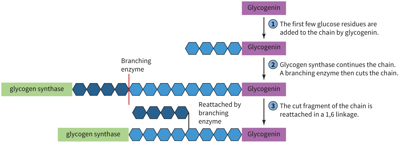
Branching enzyme transfers a 7-mer of glucose residues to the hydroxyl on the 6 carbon of a glucose residue at least four glucose residues from the branch point
Synthesis of Glycogen
Figure 8.3 Synthesis of glycogen.
Branching Enzyme
Figure 8.4 Branching enzyme.
Glycogenolysis Defined
Glycogenolysis is the breakdown of glycogen.
Is degraded one glucose at a time
Requires several important components
- Glycogen phosphorylase
- Glycogen debranching enzyme
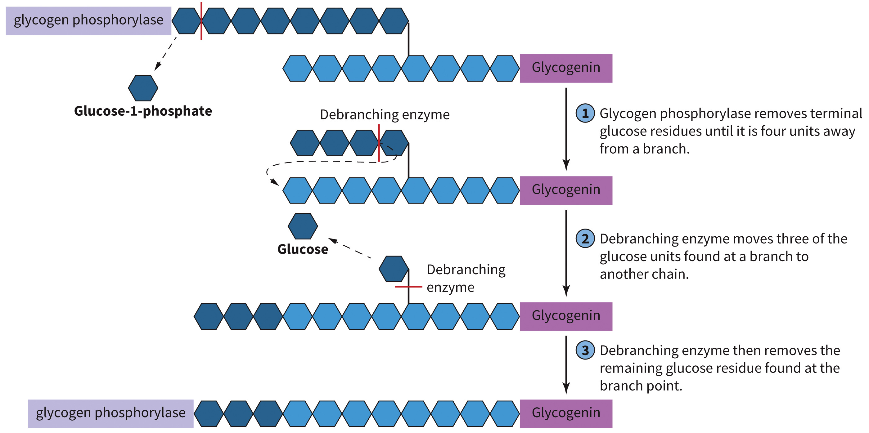
Glycogen phosphorylase removes glucose molecules at their a-1,4 linkages and requires pyridoxal phosphate as a cofactor
Glycogen debranching enzyme removes three of those four residues, transferring them to the free number 4 carbon terminus of another branch
Glycogen Breakdown
Figure 8.5 Glycogen breakdown.
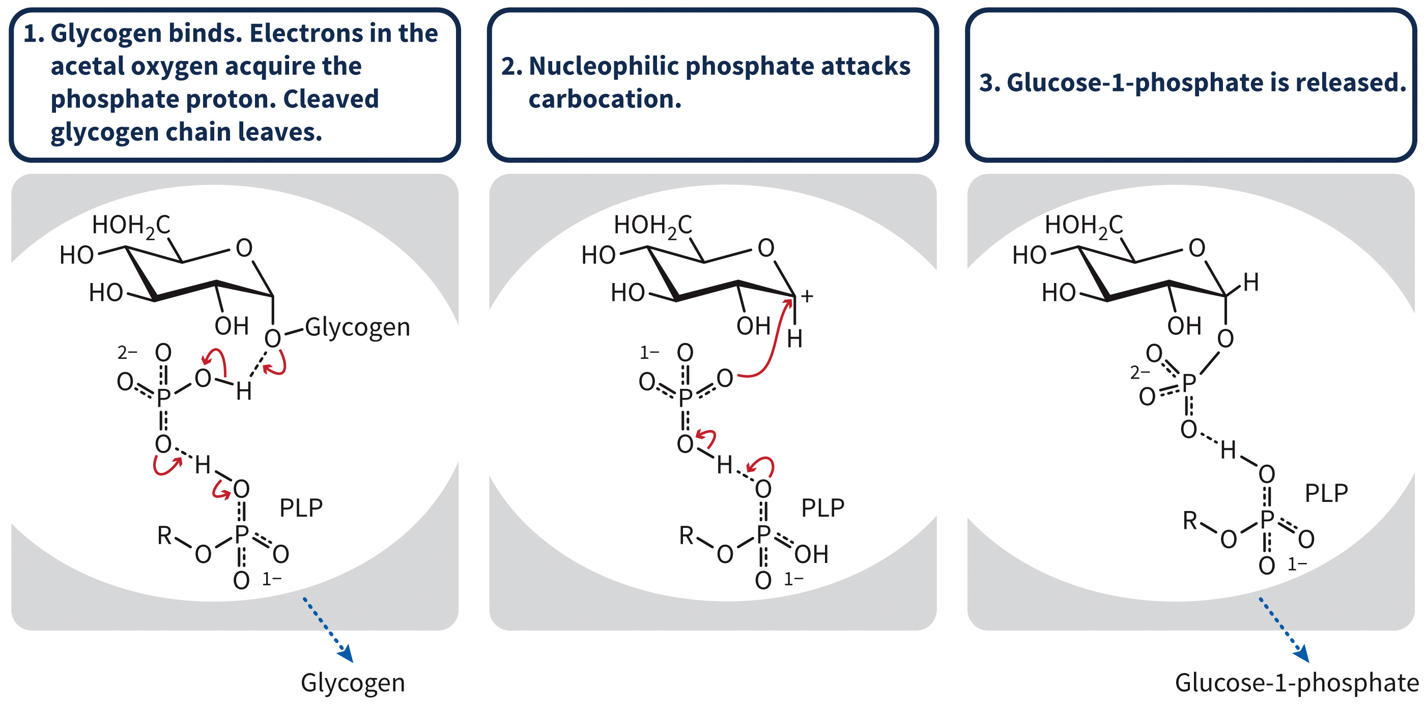
Mechanism of Glycogen Phosphorylase
Figure 8.6 Mechanism of glycogen phosphorylase.
Regulation of Glycogenesis and Glycogenolysis
Glycogen phosphorylase and glycogen synthase must be regulated.
PKA is the main regulator of both enzymes.
Kinases are regulated through signaling cascades.
Figure 8.7 Overview of the regulation of glycogen metabolism.

Allosteric Regulation of Glycogen Metabolism
ATP and glucose-6-phosphate are allosteric regulators
Glycogen synthase and glycogen phosphorylase naming convention:
- A—active
- B—less active
Phosphorylation determines whether the enzyme is active or not
Glycogen synthase and glycogen phosphorylase work in opposition
This means that glycogen synthase is most active (the a state) while dephosphorylated, but that glycogen phosphorylase is most active (the a state) when phosphorylated.
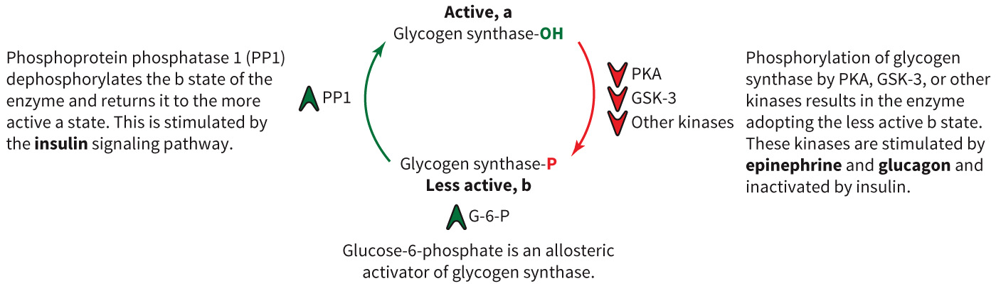
Regulation of Glycogen Synthesis
Pathways that phosphorylate glycogen synthase will inactivate it
Glycogen synthase: phosphorylated by GSK-3 and PKA
Regulated by Akt, PP1, and G-6-P
When glucose levels in the plasma are high, intracellular levels of glucose (and therefore of G-6-P) will increase, triggering the cell to store some of this G-6-P as glycogen.
Figure 8.10 Regulation of glycogen synthesis.
When glucose levels in the plasma are high, intracellular levels of glucose (and therefore of G-6-P) will increase, triggering the cell to store some of this G-6-P as glycogen.
Regulation of Glycogen Breakdown
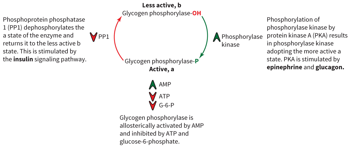
Glycogen phosphorylase is activated by phosphorylation and deactivated by dephosphorylation.
Activated by PKA and
Dephosphorylated by PP1
Figure 8.11 Regulation of glycogen phosphorylase.
G Protein-Coupled Receptors (GPCR) Cell Signaling Cascade
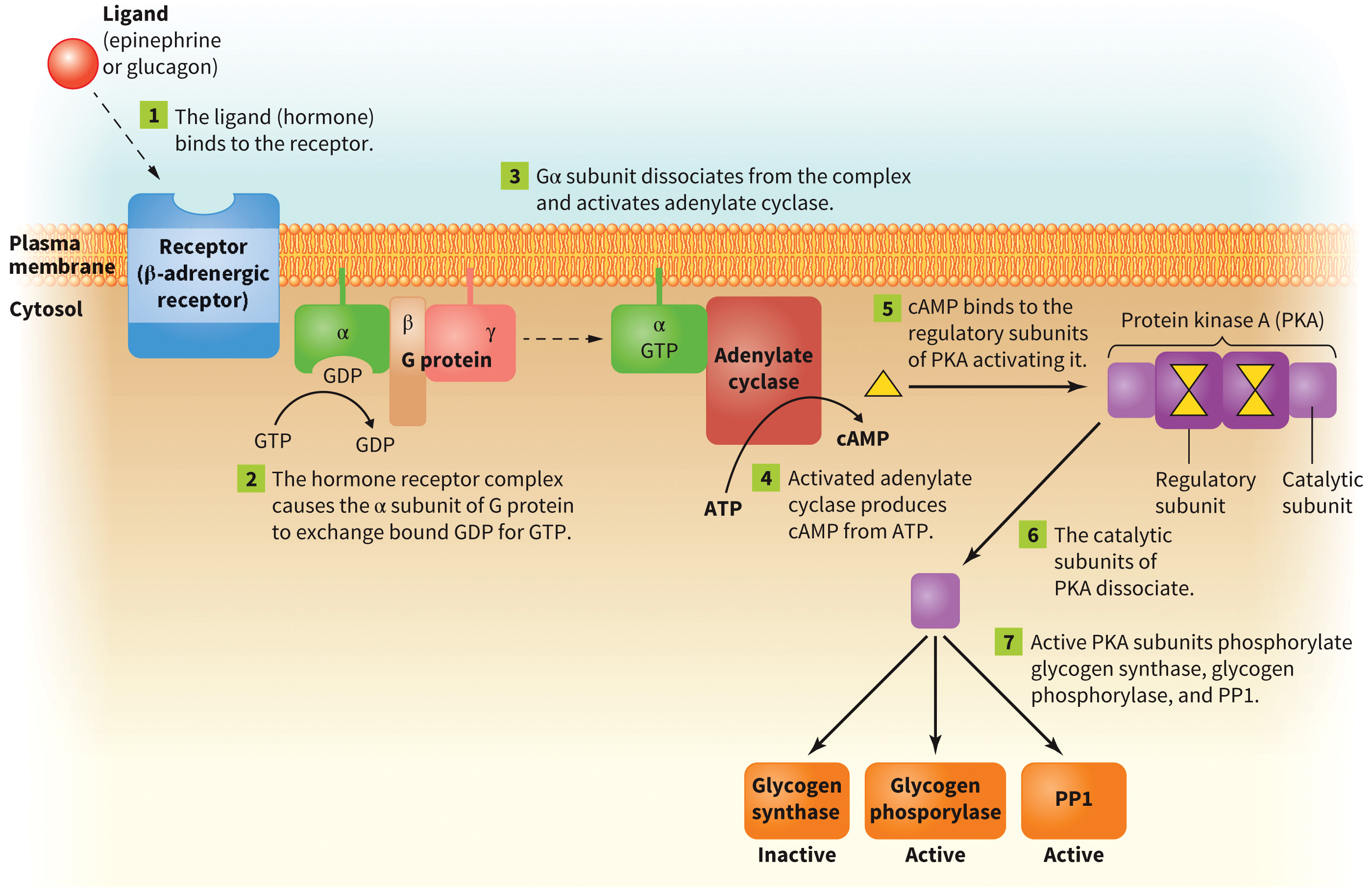
Ligand binds to receptor (G protein).
Adenylate cyclase is activated.
cAMP is generated from ATP.
cAMP = second messenger
Glycogen metabolism is dependent on PKA (cAMP dependent).
PKA has 2 regulatory and 2 catalytic subunits
Termination of signal
Tissue-specific expression of these receptors helps to fine tune and integrate whole-body glycogen metabolism
Protein Phosphatase 1 (PP1)
Enzyme anchored in the cytosol in a complex with other proteins that regulate glycogen metabolism
Is scaffolder to GM, a phosphatase that targets glycogen molecules
Regulatory proteins include proteins include glycogen phosphorylase, phosphorylase kinase and glycogen synthase.
GPCR Cell Signaling Cascade: Glucagon and Epinephrine
Figure 8.8 Action of glucagon and epinephrine on the cell.
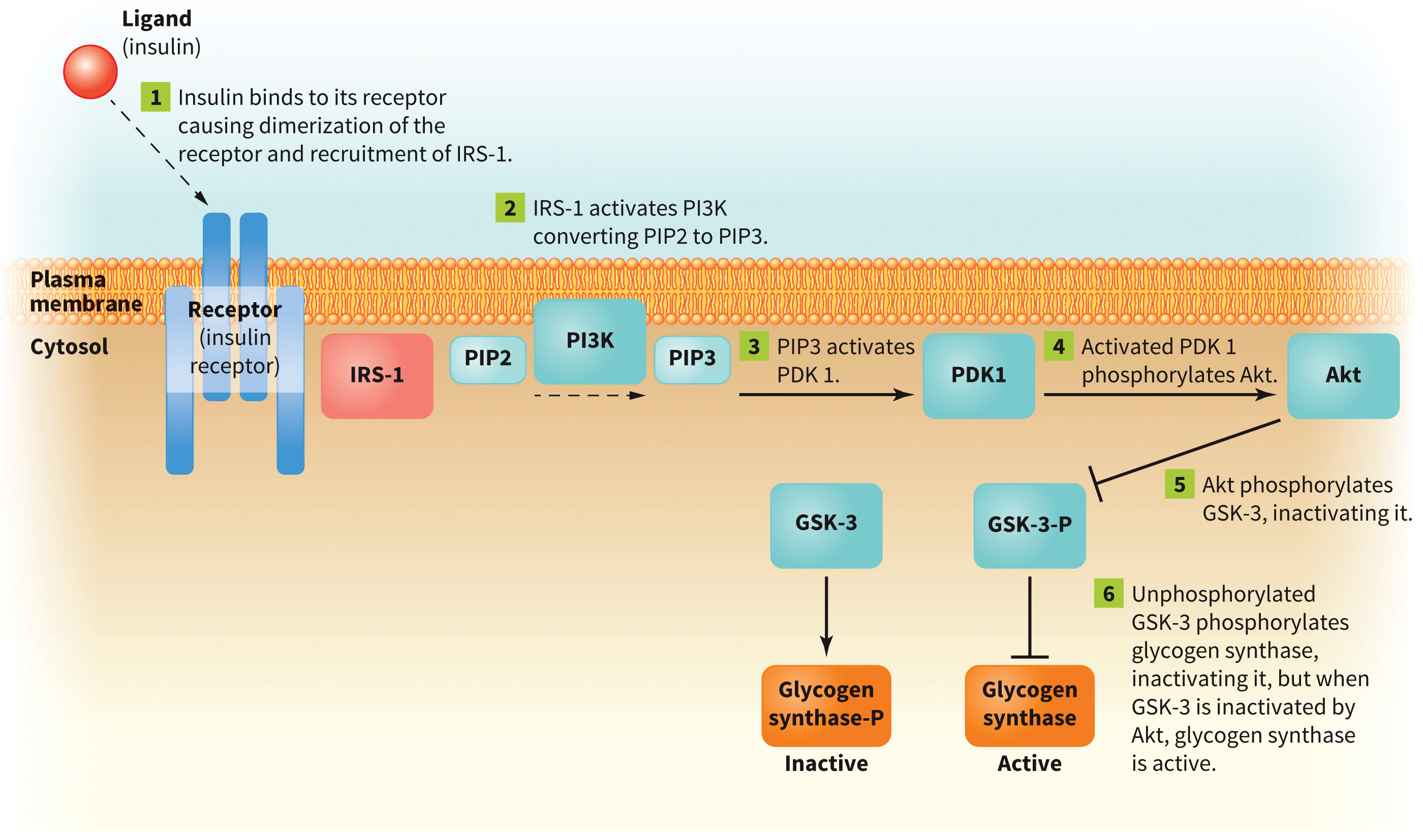
Insulin Signaling Cascade
Ligand (insulin) binds to receptor (insulin receptor).
Receptor is dimerized.
Autophosphorylation occurs.
Akt is activated.
Stimulates glycogenesis
Glycogenolysis is blocked stimulating PP1 (protein phosphatase 1)
Example of a tyrosine receptor kinase
PI3 kinase phosphorylates phosphatidyl inositol 4, 5 bisphosphate into phosphatidyl inositol 3, 4, 5 trisphosphate (initiates phosphoinositide signaling pathway) - second messenger
Insulin Signaling
Figure 8.9 Insulin signaling.
Effects of Hormones on Glucose Metabolism
| Insulin in muscle | Insulin in liver | Glucagon in muscle | Glucagon in liver | Epinephrine in muscle | Epinephrine in liver | |
|---|---|---|---|---|---|---|
| Glycogenesis | Up | Up | – | Down | Down | Down |
| Glycogenolysis | Down | Down | – | Up | Up | Up |
Section 8.2 Learning Objective
Describe the reactions of the pentose phosphate pathway.
Pentose Phosphate Pathway
Produces monosaccharides,
Also known as the pentose phosphate shunt, hexose monophosphate shunt, and phosphogluconate pathway
Is divided into two phases
- Oxidative
- Nonoxidative
Pathway occurs in the cytosol - Oxidative (produces
) - Non–oxidative (alters structure of carbohydrates)
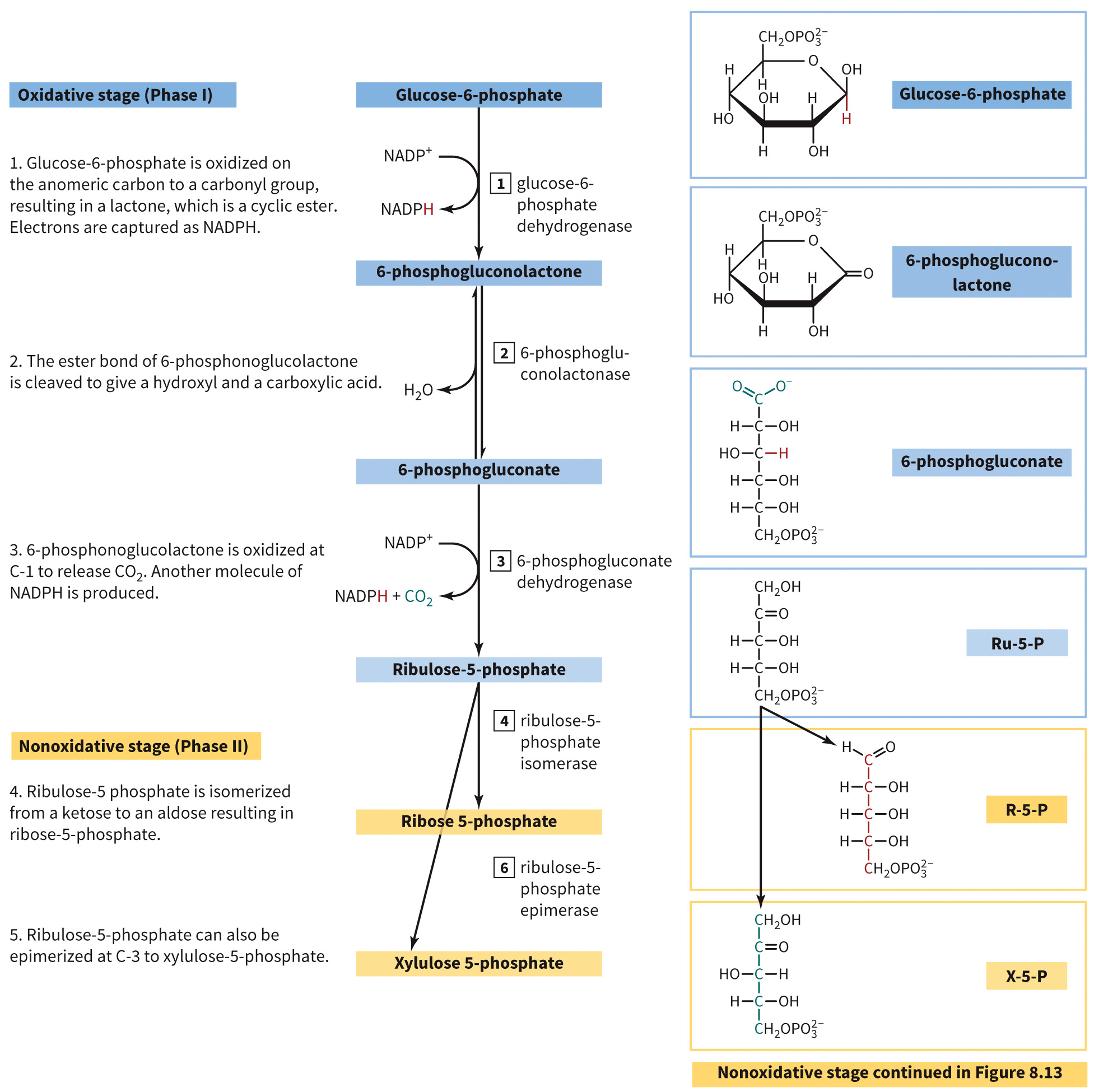
Oxidative Steps of Pentose Phosphate Pathway
Step 1 is the committed step (glucose-6-phosphate dehydrogenase).
Products made: 2 molecules of
Figure 8.12 Oxidative steps of the pentose phosphate pathway.
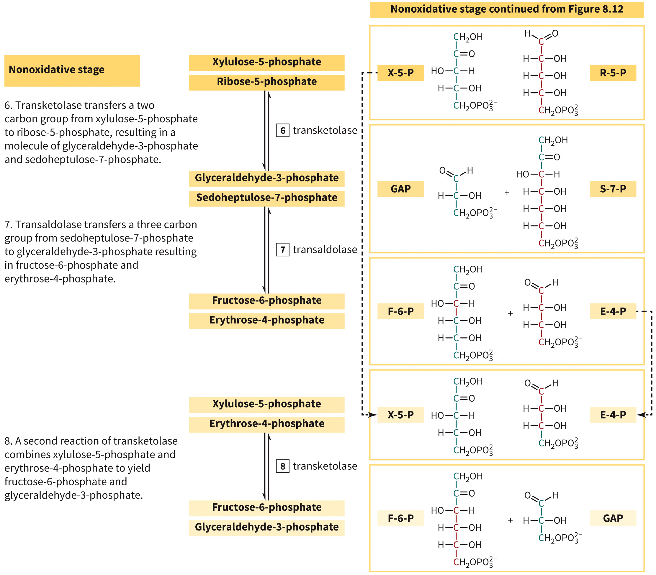
Nonoxidative Steps of Pentose Phosphate Pathway
Figure 8.13 Nonoxidative reactions of the pentose phosphate pathway.
Epimers Defined
Epimers are type of isomer that differ only in the stereochemistry about a single chiral center.
Examples include galactose (C-4 epimer) of glucose.
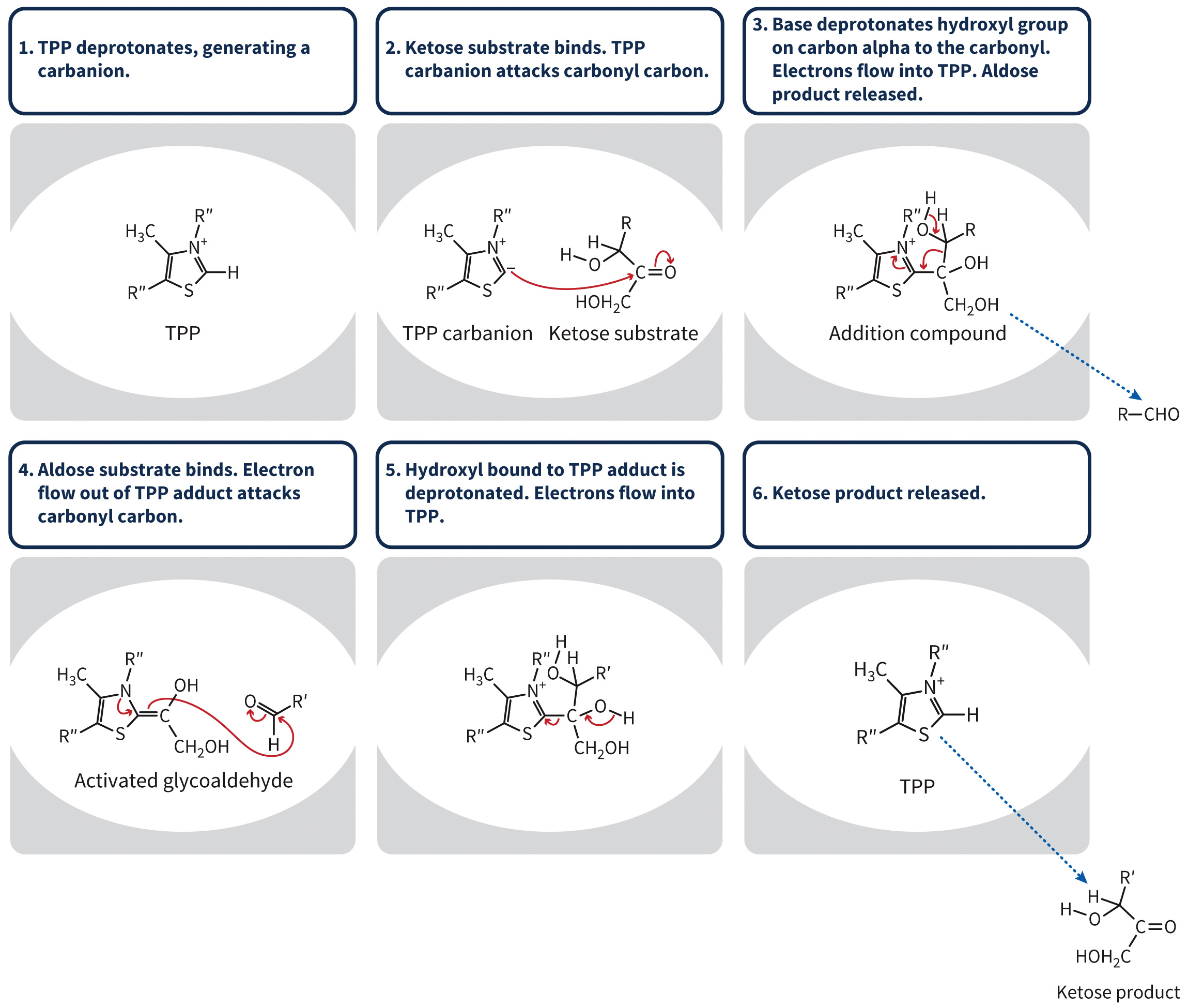
Transketolases Defined
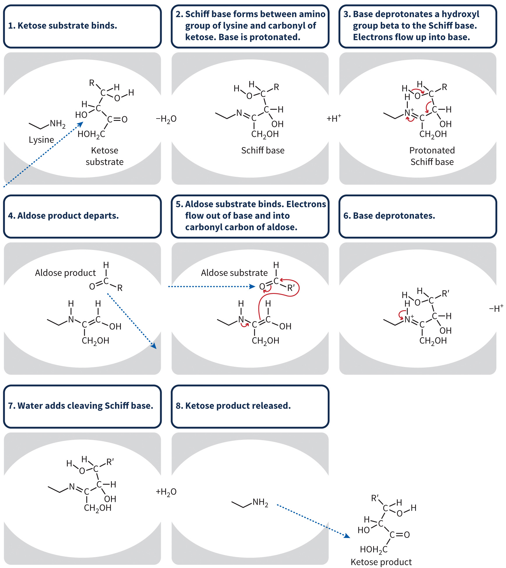
Transketolases are enzymes that use thiamine pyrophosphate (TPP) and an aldose substrate to form a ketose.
Mechanism of a Transketolase
Figure 8.14 Mechanism of transketolase.
Transaldolases
Transaldolases are enzymes that use an active lysine to form a Schiff base with an aldose substrate and a carbonyl carbon to form a ketose.
Mechanism of a Transaldolase
Figure 8.15 Mechanism of transaldolase.
Regulation of the Pentose Phosphate Pathway
Glucose-6-phosphate dehydrogenase is regulated by levels of
Monosaccharides (i.e., ribose) can be converted to hexoses for entry into glycolysis.
Carbohydrates can be oxidized into
Occurs in the cytosol with glycolysis and TCA cycle
Glutathione and the Pentose Phosphate Pathway
Glutathione is a tripeptide antioxidant that neutralizes reactive oxygen species.
Can be oxidized to a dimer via glutathione peroxidase
Glutathione dehydrogenase uses
Glucose-6-Phosphate Dehydrogenase Deficiency
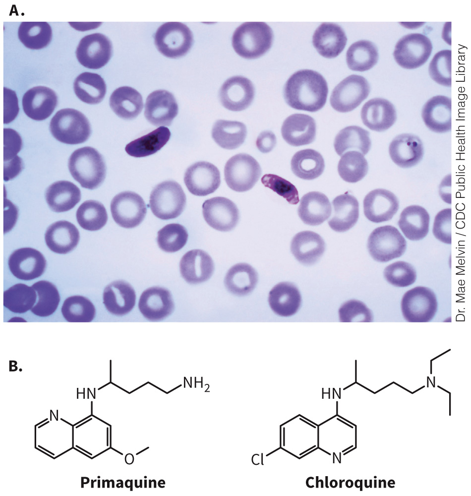
Partial deficiency can be advantageous.
Provides resistance to malaria
Figure 8.16 Primaquine and chloroquine.
400 million persons have this genetic mutation; most are asymptomatic
Can potentiate effects of antimalarial drugs
Fava beans and favism
Carbohydrate Response Element-Binding Protein (ChREBP)
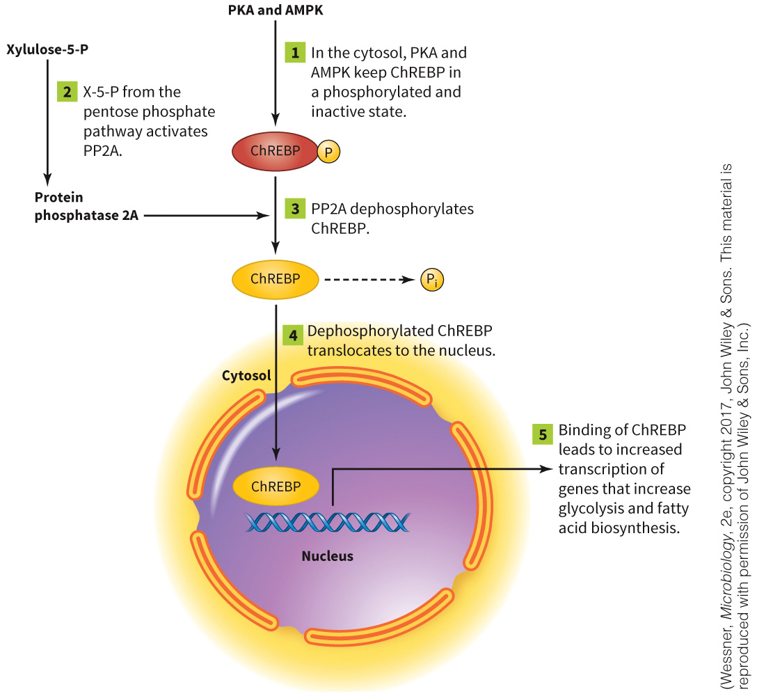
A transcription factor that responds to high levels of carbohydrates
Regulates genes involved in carbohydrate and lipid metabolism
Xyulose-5-phosphate actives PP2A, which activates ChREBP.
ChREBP increases expression of genes involved in energy storage.
ChREBP Mechanism
Figure 8.18 Carbohydrate response element binding protein (ChREBP).
Section 8.3 Learning Objective
Compare different types of glycosolyated molecules (glycoproteins, glycolipids, proteoglycans, and peptidoglycans) with regard to structure and function.
Glycoproteins Defined
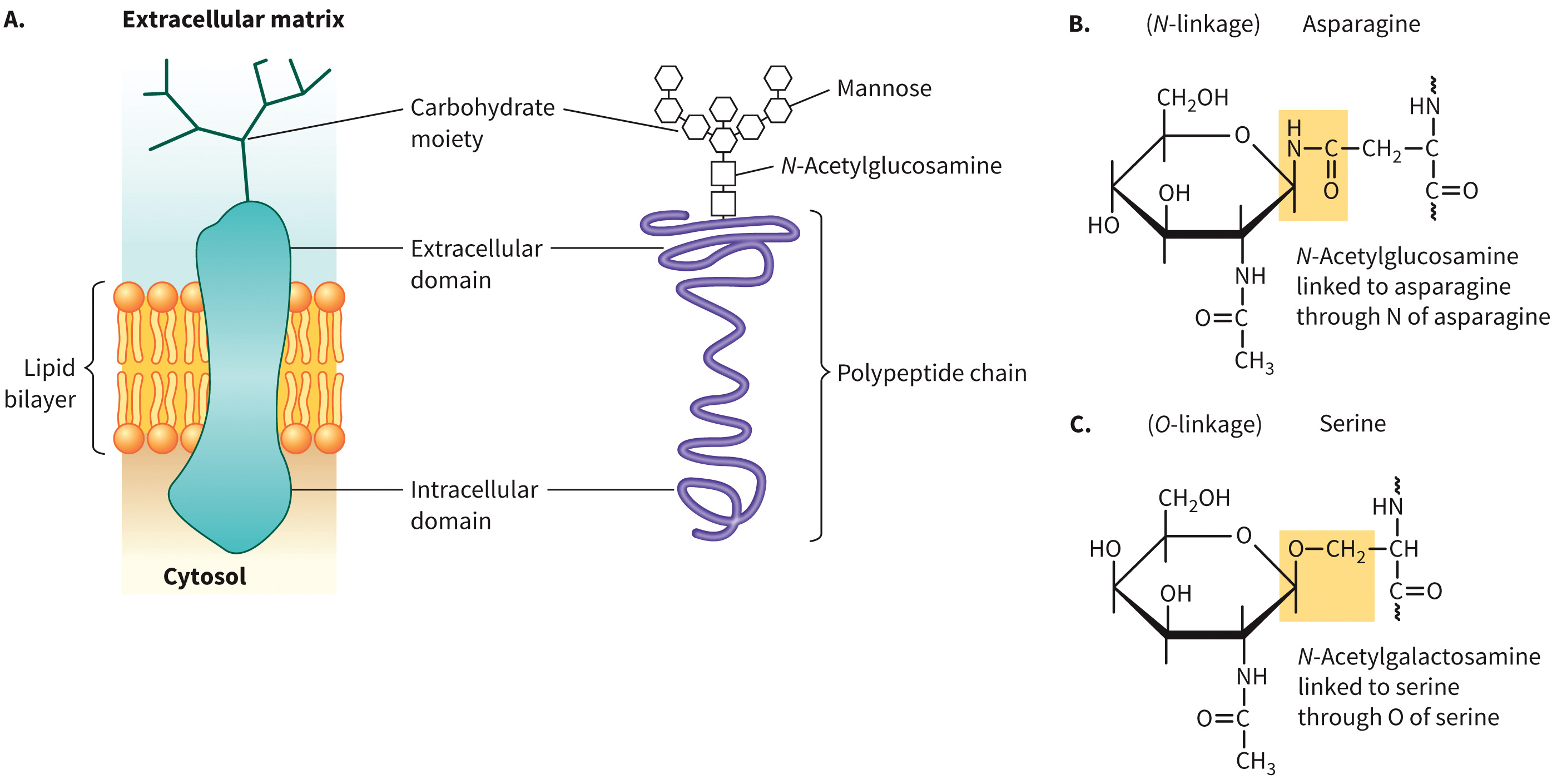
Glycoproteins are membrane-bound or extracellular proteins with some amount of carbohydrate modification.
Can be either N-linked (amide) or O-linked (acetal)
Can regulate enzyme activity, protein stability, and play a role in protein folding and protein trafficking
Examples include amino sugars, derivatives of sialic acid, and ABNO blood groups.
Modifications are made initially in the endoplasmic reticulum, and are then continued in the Golgi complex
Glycoprotein Example
Figure 8.19 An example of a glycoprotein.
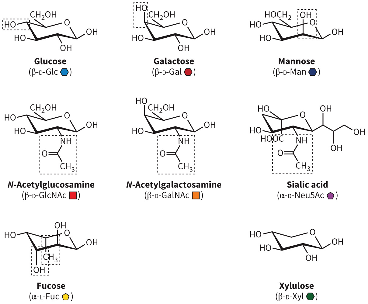
Common Carbohydrates Found in Glycoproteins
Figure 8.20 Common carbohydrates found in glycoproteins.
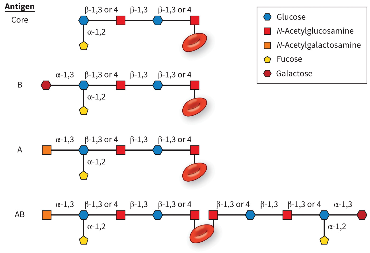
ABO Blood Group Antigens
O—universal donor
AB—universal recipient
Blood group compatibility
Note that + or - is based on the Rhesus factor, indicating the presence or absence of the protein
Figure 8.21 ABO blood group antigens.
Glycolipids Defined
Glycolipids are membrane phospholipids with an attached carbohydrate moiety exposed to the external environment.
Amphipathic molecules that contain either a glycerol or sphingosine backbone
Four examples include:
- lipopolysaccharides
- glycosylphosphatidylinositol (GPI)
- cerebrosides
- gangliosides
Generally found on the outer leaflet of the plasma membrane
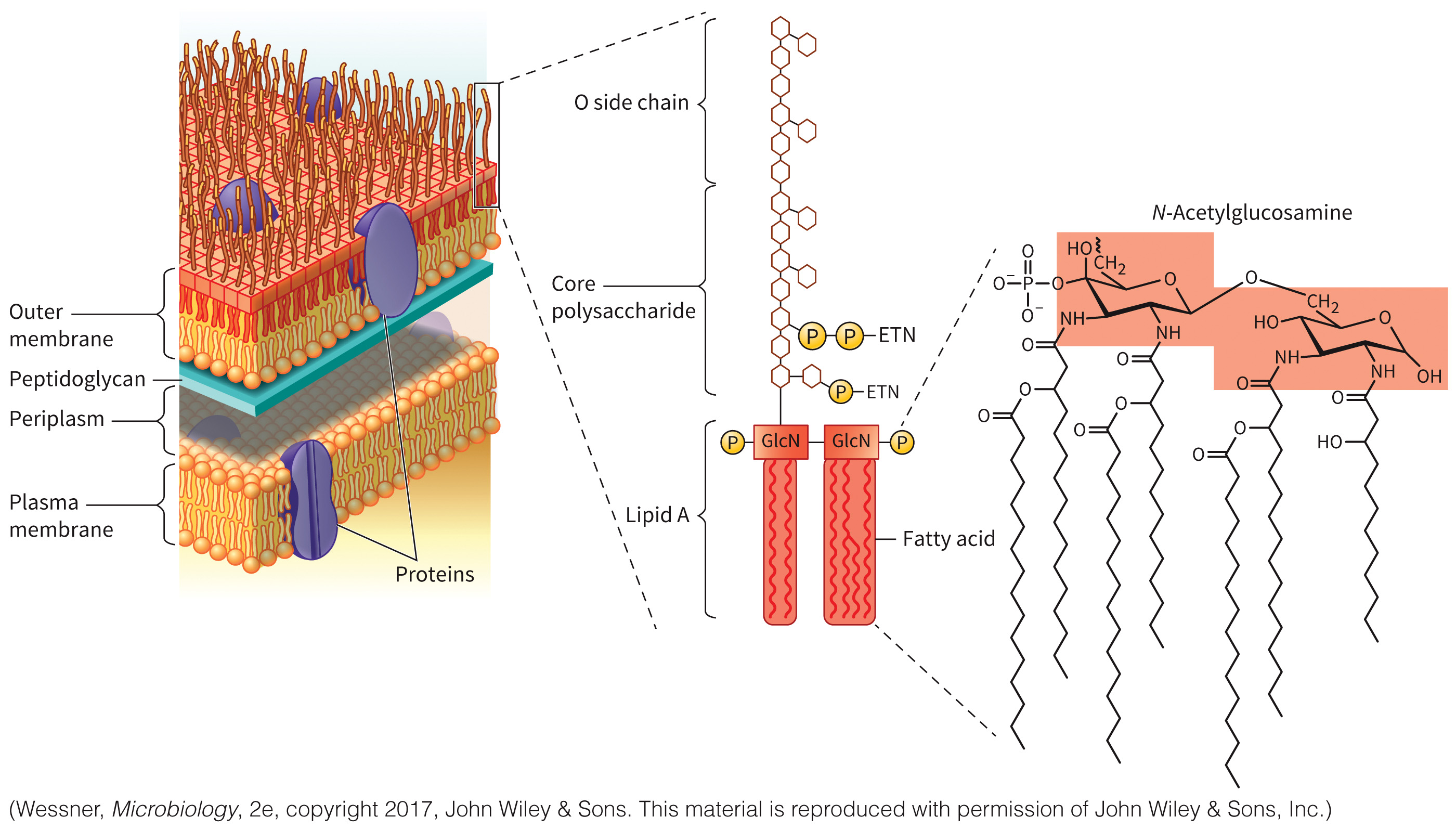
Lipopolysaccharide
Component of the bacterial outer membrane
Is a pyrogen and toxic in animals – can lead to septic shock seen in advanced infections
Three antigens - the O antigen, a core oligosaccharide, and lipid A
Figure 8.22 Structure of lipopolysaccharide.
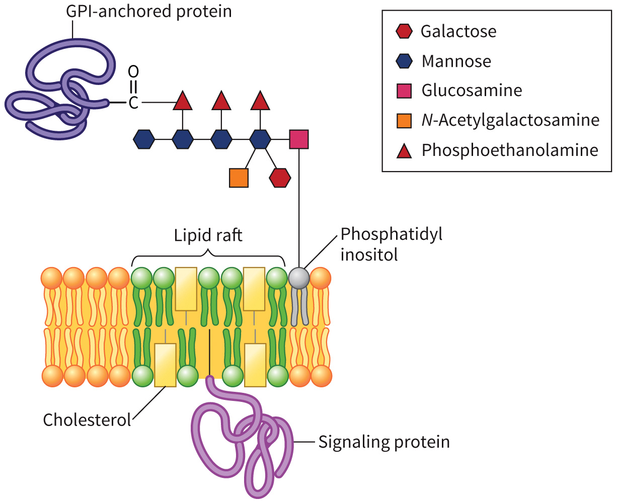
Glycosylphosphatidylinositol (GPI)
Anchor proteins to the outer leaflet of the plasma membrane
Used by several signal transduction pathways to localize proteins in the plasma membrane
Figure 8.23 Structure of a glycosyl phosphoinositide (GPI) anchor.
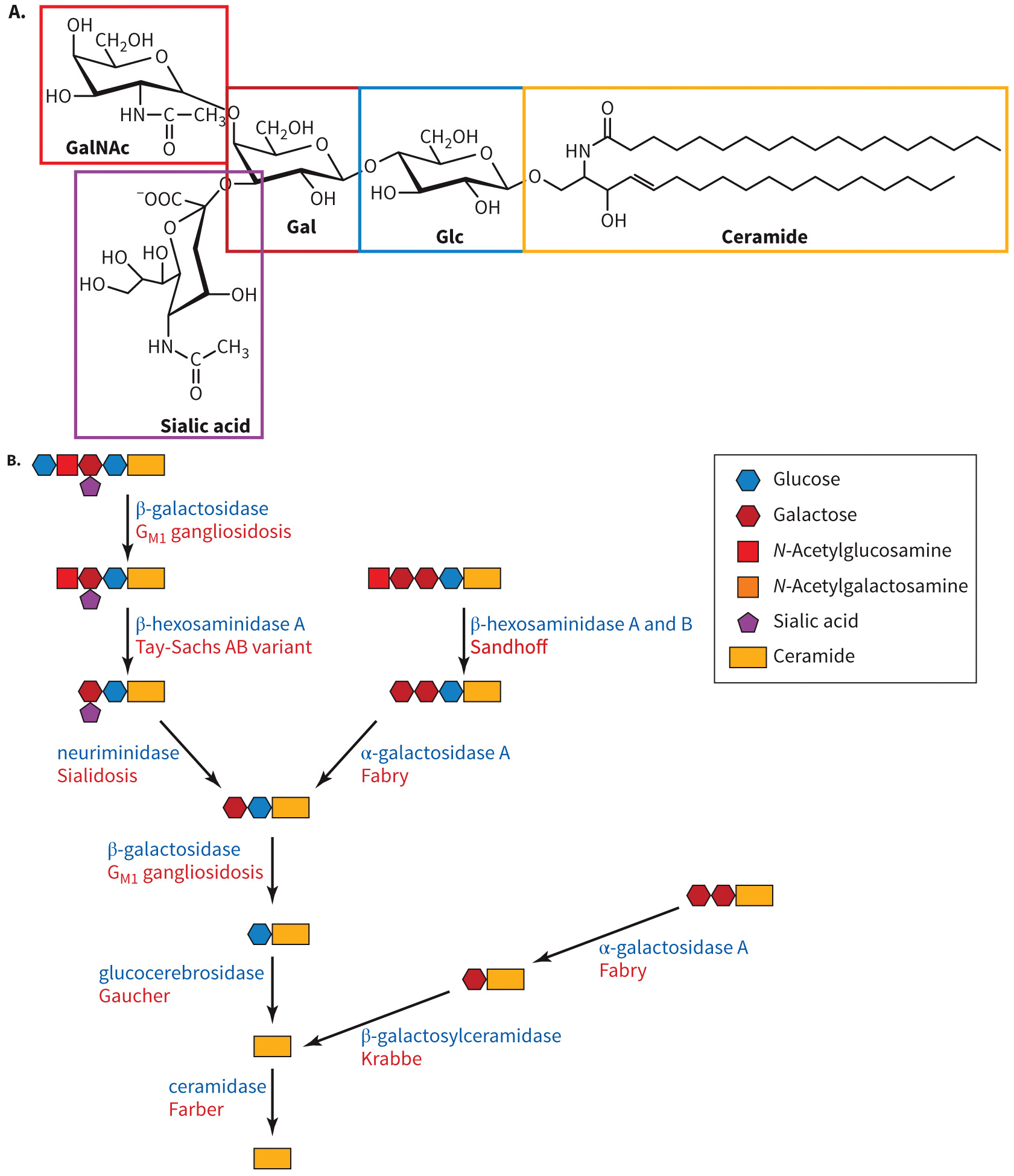
Cerebrosides
Glycosylated sphingolipids that have a single sugar attached to a free OH group through a ceramide through a glycoside linkage
Are internalized and degraded by lysosomes
Figure 8.24 Structures of cerebrosides and gangliosides.
Gangliosides
Glycosylated sphingolipids that have three to seven monosaccharides attached
Are internalized and degraded by lysosomes
Proteoglycans Defined
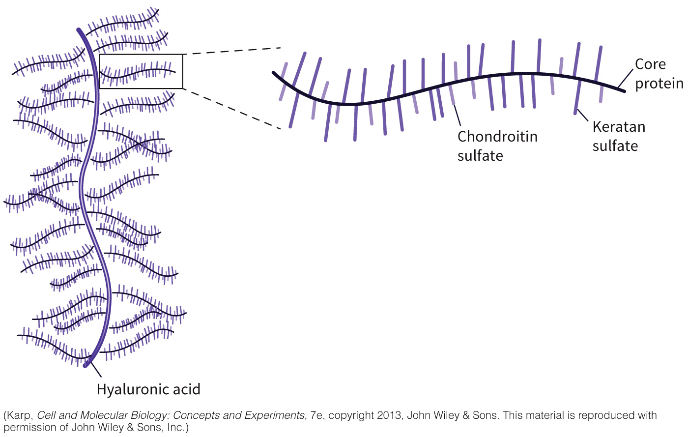
Proteoglycans are extensive mesh nets of polysaccharides joined to fibrous proteins
Major component of the extracellular matrix
Contain a glycosaminoglycan and a protein core
Have more carbohydrate nature
Most are viscous (act as gels/lubricants)
Structure of a Proteoglycan
Figure 8.26 Structure of a proteoglycan.
Examples of Glycosaminoglycans
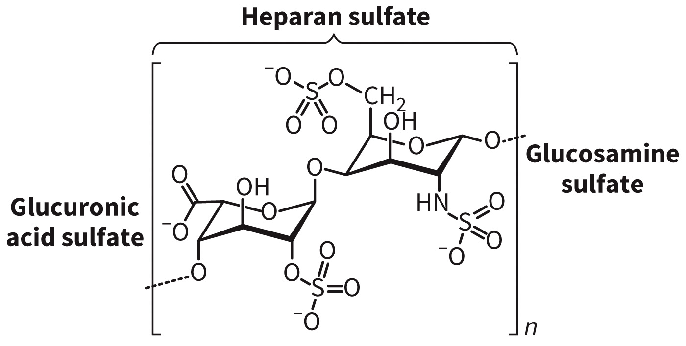
- Heparan sulfate
- Found in extracellular matrices of most animals
- Linear polysaccharide comprises repeating disaccharides, usually glucuronic acid and N-acetylglucosamine
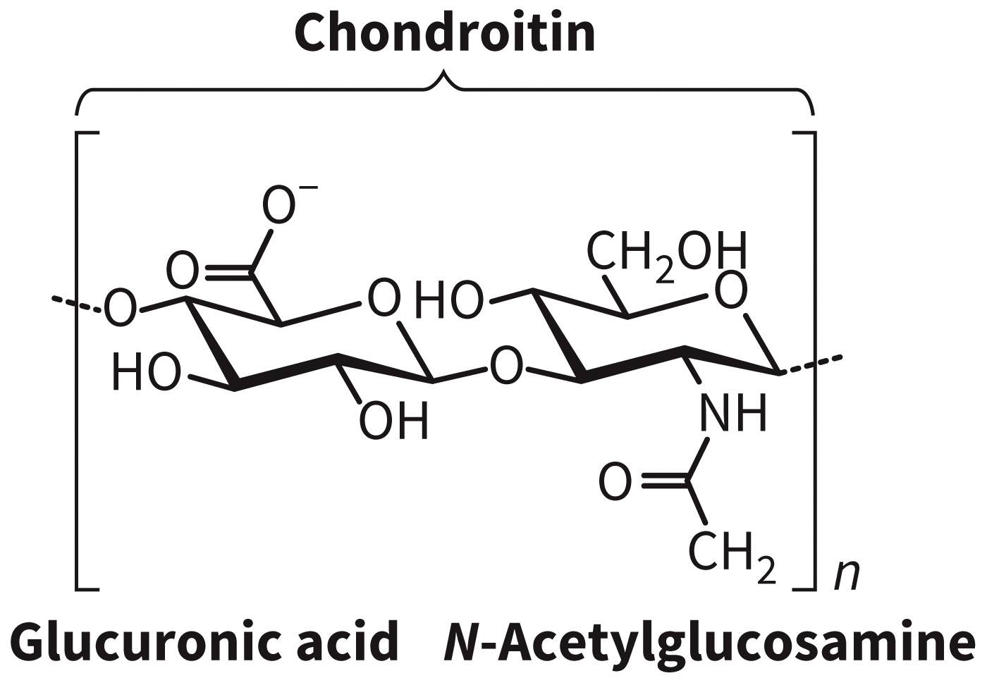
- Chondroitin sulfate
- Found in tendons, cartilage
- Linear polysaccharide comprises repeating glucuronic acid and N-acetylgalactosamine disaccharides
Examples of Glycosaminoglycans
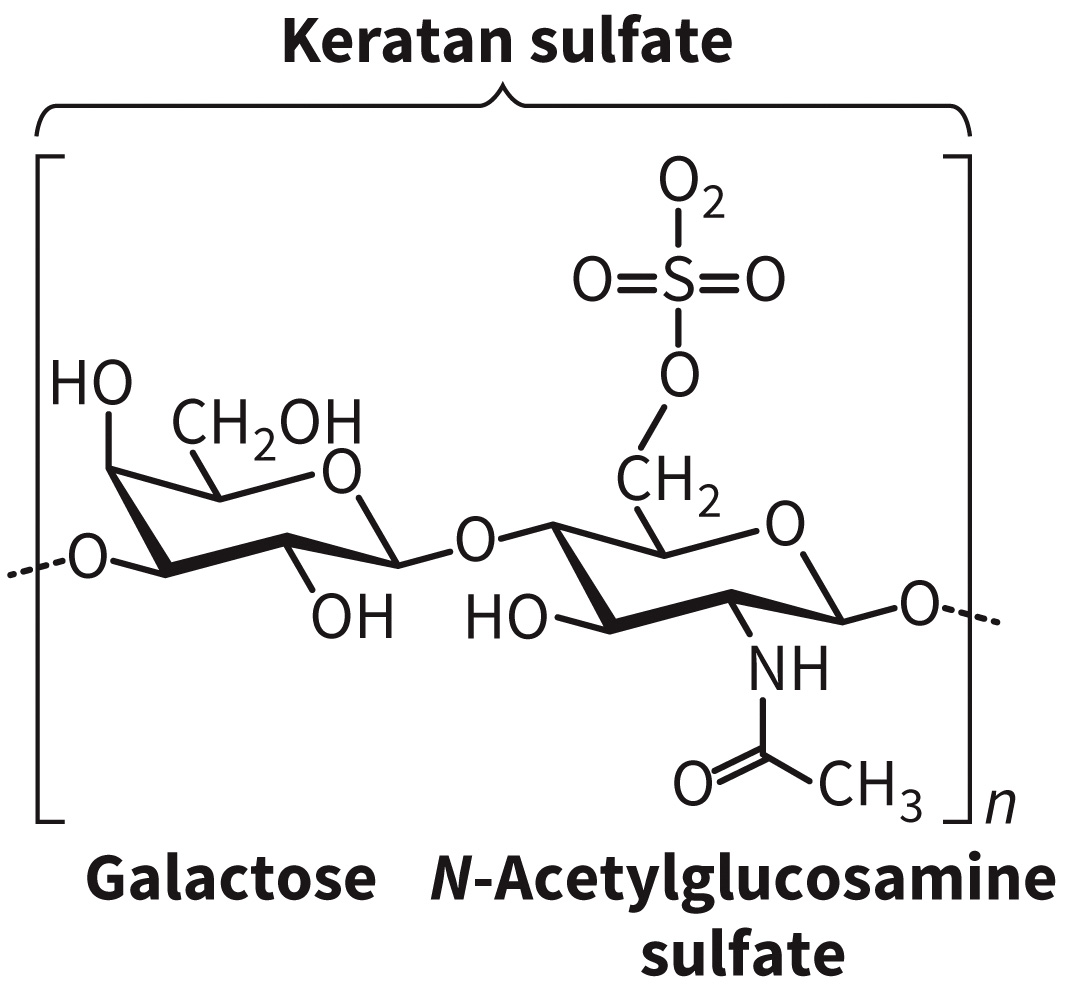
- Keratan sulfate
- Found in bone, horn, and cornea
- Linear polysaccharide is a polymer of galactose and N-acetylglucosamine
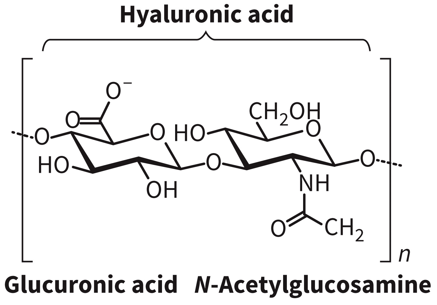
- Hyaluronic acid
- Non-proteoglycan matrix
- Secreted as a lengthy linear polysaccharide of glucuronic acid and N-acetylglucosamine
- Synthesized by a complex in the plasma membrane
Peptidoglycans Defined
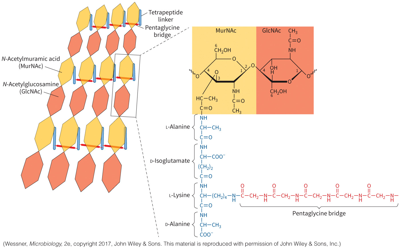
Peptidoglycans are lengthy chains of polysaccharides cross-linked by peptides and found in bacterial cell walls.
Composed of a dimeric repeat of N-acetylglucosamine (GlcNAc) and N-acetylmuramic acid (MurNAc) in a β-1,4 linkage
Antibiotics can block cell wall formation.
Structure of the Peptidoglycan Cell Wall in Gram-Positive Bacteria
Figure 8.27 Structure of the peptidoglycan cell wall in gram-positive bacteria.
Section 8.4 Learning Objective
Analyze how extracellular matrices and biofilms are synthesized and function.
Extracellular Matrix Defined
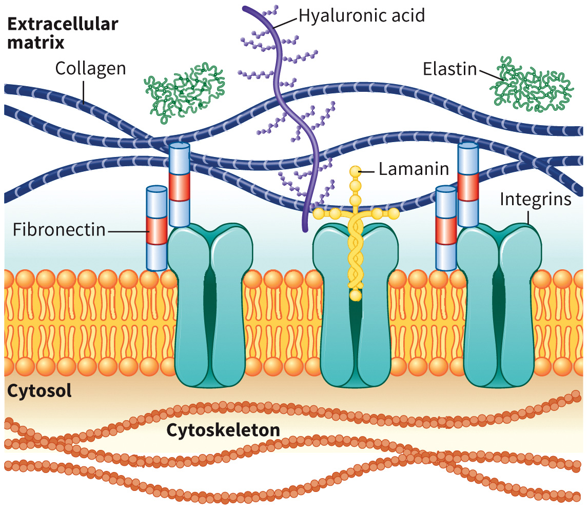
Extracellular matrix is a tissue made of proteins, glycoproteins, and proteoglycans in a fibrous, gel-like mesh.
Some proteins play a role in ligand binding, immune response, and regulation of growth and development.
Figure 8.29 Schematic view of the extracellular matrix. Several fibrous proteins are included: collagen, elastin, fibronectin, and laminin.
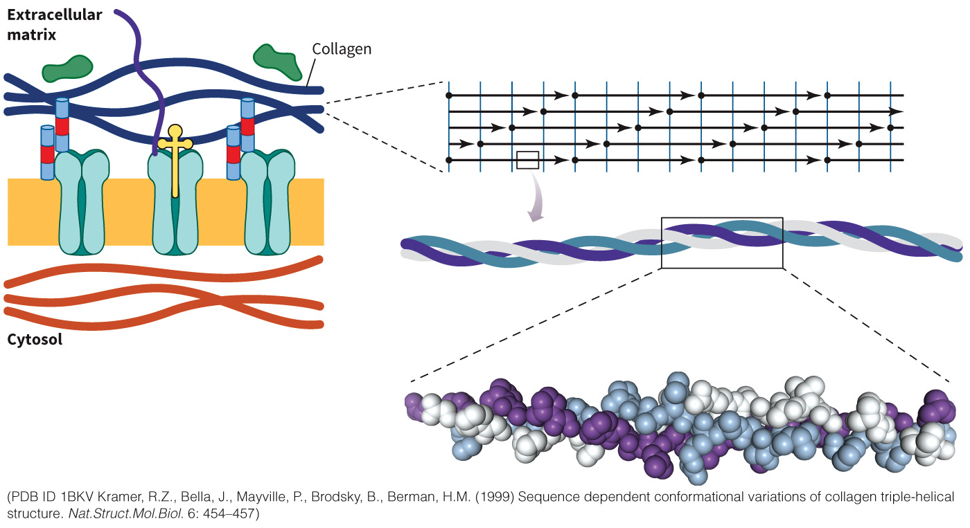
Collagen
Protein found in high amounts in the extracellular matrix
Adopts a triple helix composed of Gly, Pro, and Lys
25% of the protein in the body (by mass) is collagen
Figure 8.30 Structure of collagen.
Collagen Synthesis
Requires Vitamin C
Proline hydroxylation is key to this process.
Succinate can enter the TCA cycle

Elastin
Responsible for the core of elastic fibers found in skin and arteries
Composed of largely hydrophobic residues
Enriched with Pro and Gly and rich in repeated stretches of Ala and Lys
Proline hydroxylation is key
Figure 8.31 Structure of elastin and the desmosine cross-link.

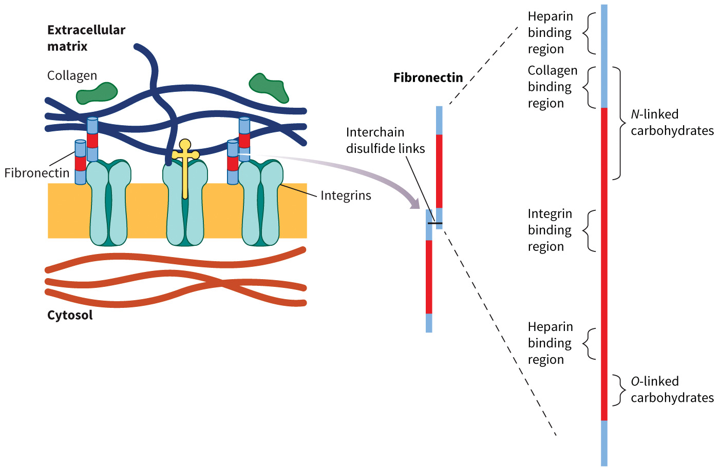
Fibronectin
Found in the extracellular matrix in a soluble form in the circulation
Functions in the clotting cascade
Acts as an adapter between the cell and the matrix
Structure of Fibronectin
Figure 8.33 Structure of fibronectin.
Laminins
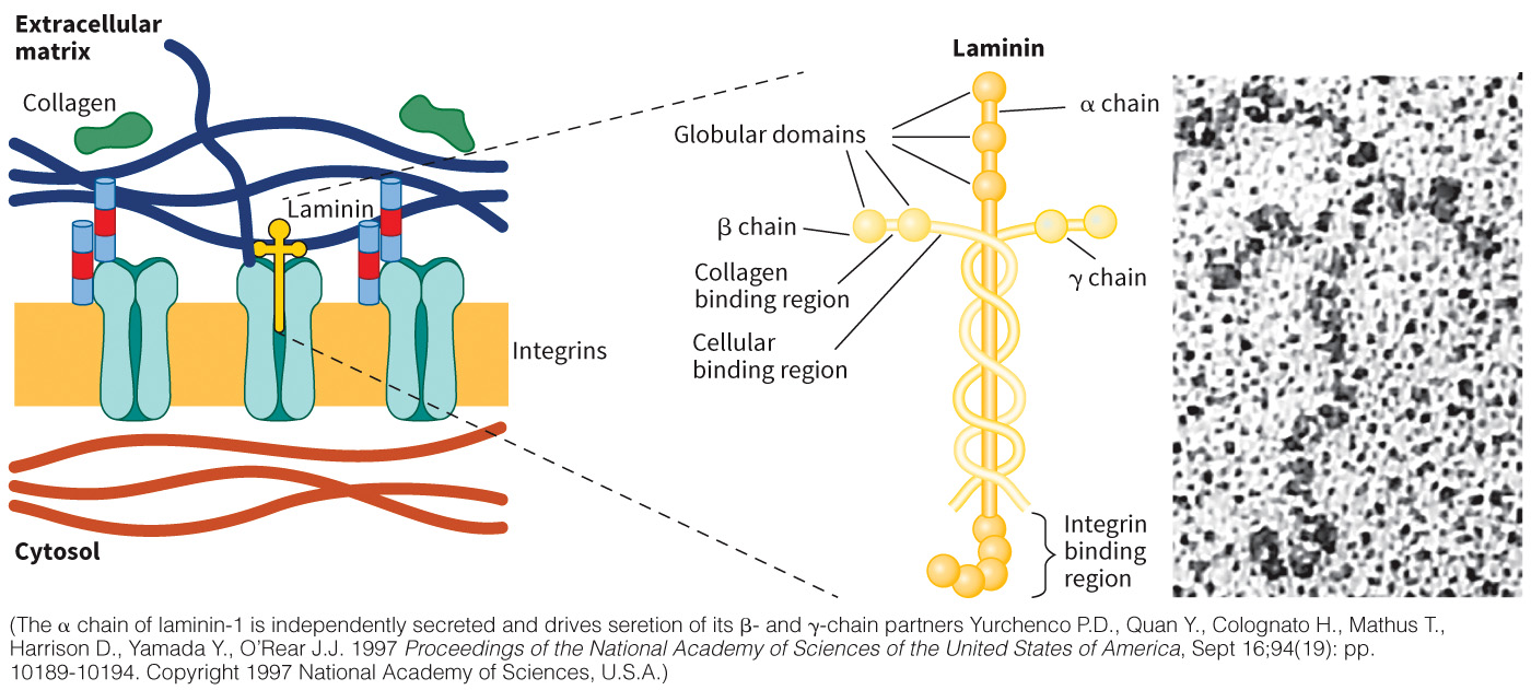
A family of proteins that function in the basal lamina, a layer of the basement membrane, a fibrous layer of connective tissue found under the epithelial layers.
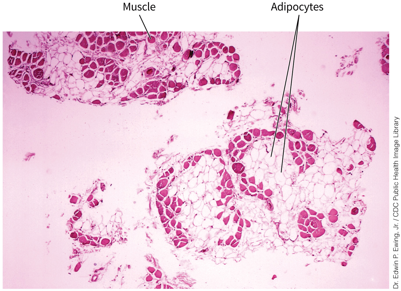
Implicated in several diseases including muscular dystrophy
Figure 8.34 Structure of the laminins.
Laminin Dysfunction and Muscular Dystrophy
Figure 8.35 Dysfunction of the laminins results in muscular dystrophy.
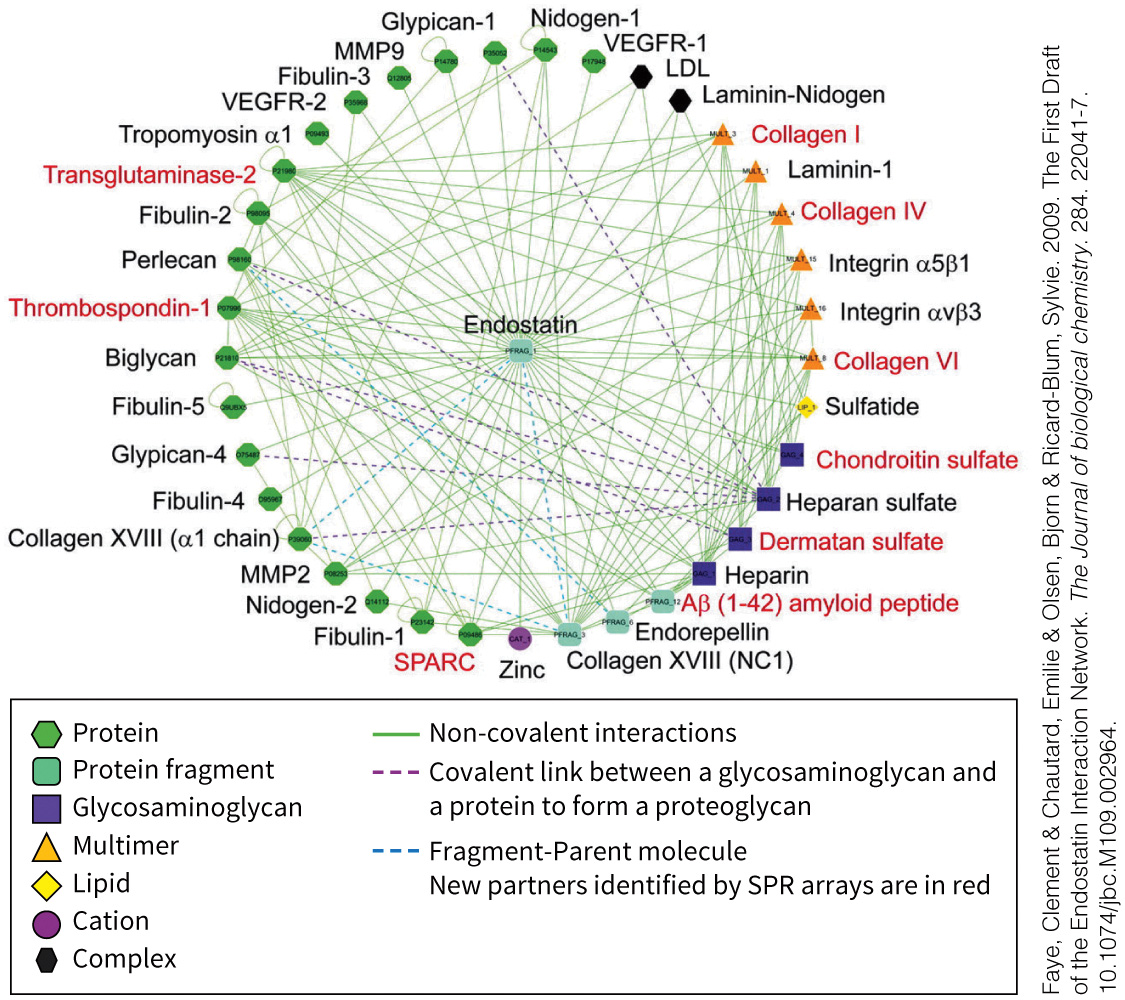
Interactome Defined
Interactome describes how large matrix proteins interact with other macromolecules.
Figure 8.36 Interactions of matrix proteins can be studied in silico.
Cell Culture Defined
Cell culture is a process in which cells are grown on a plastic tissue culture dish for researchers to study cells under different conditions.
Cells are grown to confluence and a chemotherapeutic agent can be added to stop cells from dividing
Figure 8.37A Cultured cells.
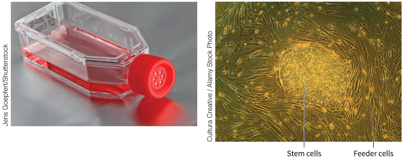
Tissue Engineering Defined
Tissue engineering describes the growth of cultured cells in a three-dimensional matrix
The scaffold, the matrix to which the cells attach, needs to provide structure for the cells to grow
Have seen promising successes with skin and vascular tissue grafts and artificial bladders
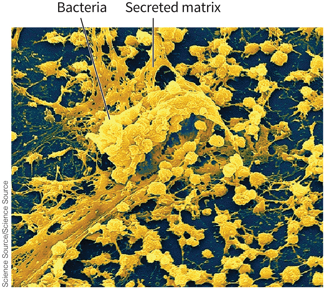
Biofilms Defined
Biofilms are associations of microbes living in a secreted matrix.
Are often found at an air–liquid or solid–liquid interface
Can be of a single species or multiple species
Examples include pond slime, dental plaque
Bacteria are often more antibiotic resistant in a biofilm.
Figure 8.39 Structure of a biofilm.
Biofilm Development
Figure 8.40 Life cycle of a biofilm.

Biofilm Decontamination and Prevention
Not simple because of surface attachment
Treatment strategies include
- blasting with a jet of oxygen–helium plasma
- ozone
Prevention strategies include - adding coating to discourage bacterial settling
Prevention additives include: antibiotics, hydrophobic oils, nanoparticles of silver, or positively charged groups such as N-alkylpyridinium salts
Biofilms in Human Health
Pseudomonas aeruginosa
- Participates in opportunistic infections
- Can be treated with antibiotics
Dental plaque - Biofilm composed of microbes (mostly Streptococcus) that live on the surface of teeth and gingival, or gum, tissues
- Can be controlled by proper dental hygiene