06 The Common Catabolic Pathway Citric Acid Cycle, the Electron Transport Chain, and ATP Biosynthesis
Outline
7.1 The citric acid cycle
7.2 The electron transport chain
7.3 ATP biosynthesis
Learning Objectives
- Describe how acetyl-CoA is oxidized and other molecules are transformed in the citric acid cycle.
- Illustrate how electron transport results in protons (
) getting pumped out of the mitochondrial matrix, and the formation of water. - Describe how the cell uses the electrochemical gradient produced in electron transport to synthesize ATP.
Section 7.1 Learning Objective
Describe how acetyl-CoA is oxidized and other molecules are transformed in the citric acid cycle.
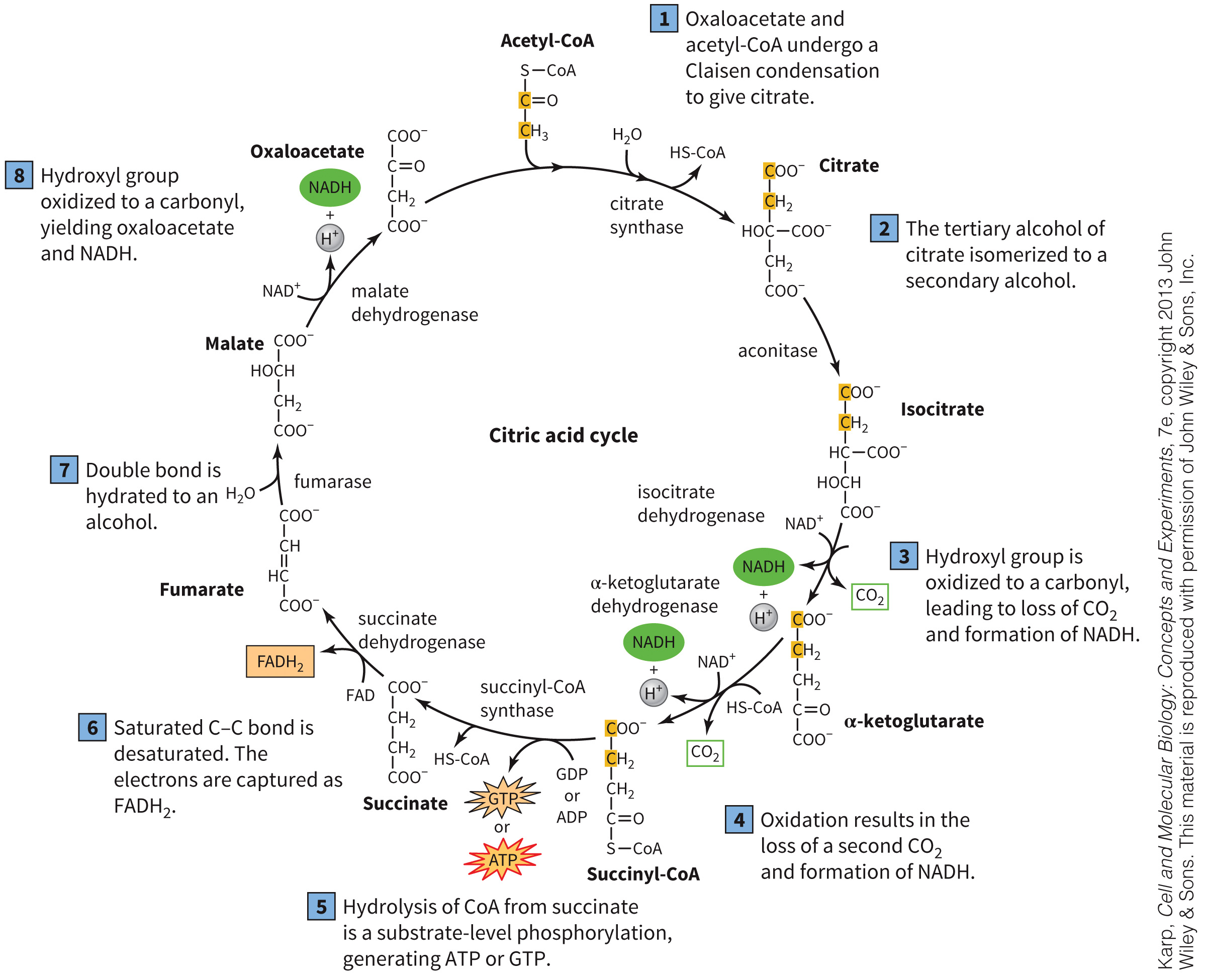
The Citric Acid Cycle
Also known as the Krebs cycle and tricarboxylic acid cycle
Eight reactions that serve as a central metabolic hub
Generates the following:
- 2
- 1 GTP or ATP
- 3
- 1
Example of a cyclic pathway
Electrons asand for use in electron transport
1= 2.5 ATP and 1 = 1.5 ATP
The Citric Acid Cycle
Figure 7.2 Citric acid cycle.
Example of a metabolon
Metabolon Defined
Metabolon is a group of enzymes performing reactions with a common purpose.
Enzymes can be localized in an organelle or part of an organelle.
Associated with glycolysis, glycogenolysis, fatty acid biosynthesis, and the electron transport
Substrate channeling may occur.
Substrate Channeling Defined
Substrate channeling is the diversion of the product of one enzymatic reaction directly into a subsequent reaction, to increase reaction rate and efficiency.
Acetyl-CoA
Important intermediate in several pathways
Product of fatty acid catabolism
Chemical bonds are energetically rich.
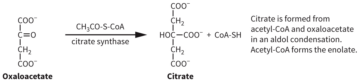
Citrate Synthase
Condensation of acetyl CoA with oxaloacetate
Produces citrate
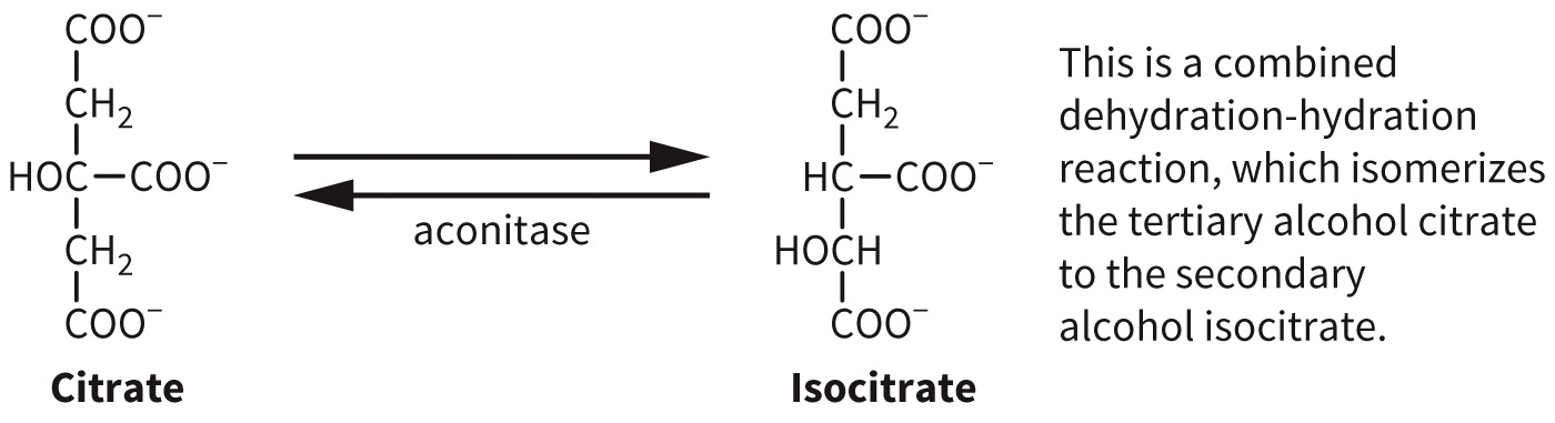
Aconitase
Isomerization of citrate to isocitrate
Reversible reaction
Produces citrate
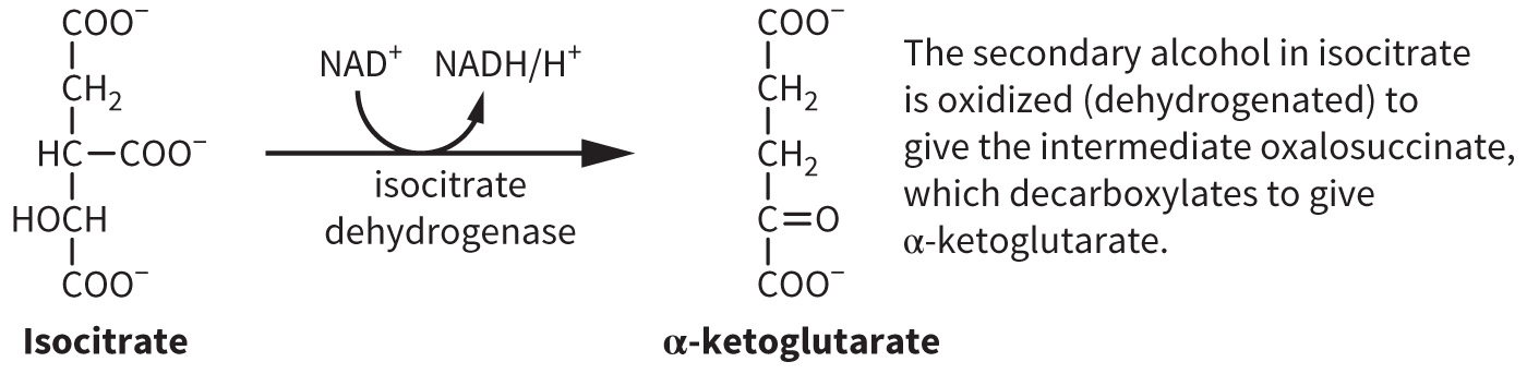
Isocitrate Dehydrogenase
Oxidation of isocitrate to α-ketoglutarate
^+$+ is reduced.
Produces citrate
Please point of the for the TCA cycle:
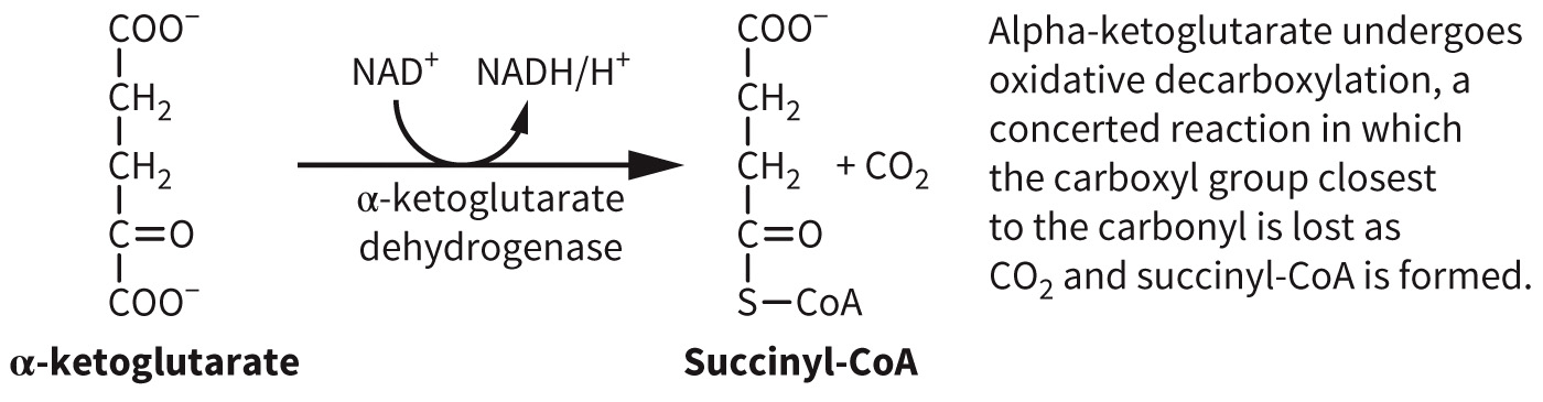
α-Ketoglutarate Dehydrogenase
α-ketoglutarate is oxidized to succinyl-CoA.
Rate-determining step
^+$+ is reduced.
α-Ketoglutarate Dehydrogenase Complex
Multienzyme complex
Three different subunits:
- E1—TPP decarboxylase
- E2—dihydrolipoyl transferase
- E3—dihidyrolipoyl dehydrogenase
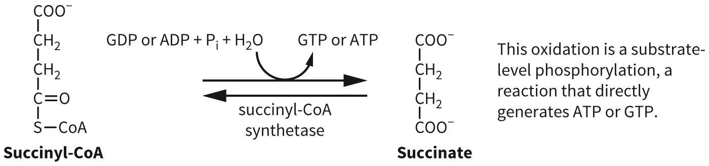
Succinyl CoA Synthetase
Substrate-level phosphorylation
- Nucleotide is generated directly by phosphorylating a nucleoside
Reversible reaction
Produces succinate and ATP or GTP
Two isoforms of enzyme – one form will produce GTP and the other will form ATP.
Succinate Dehydrogenase
Oxidation of succinate
FAD is reduced.
Produces fumarate, a trans-dioic acid
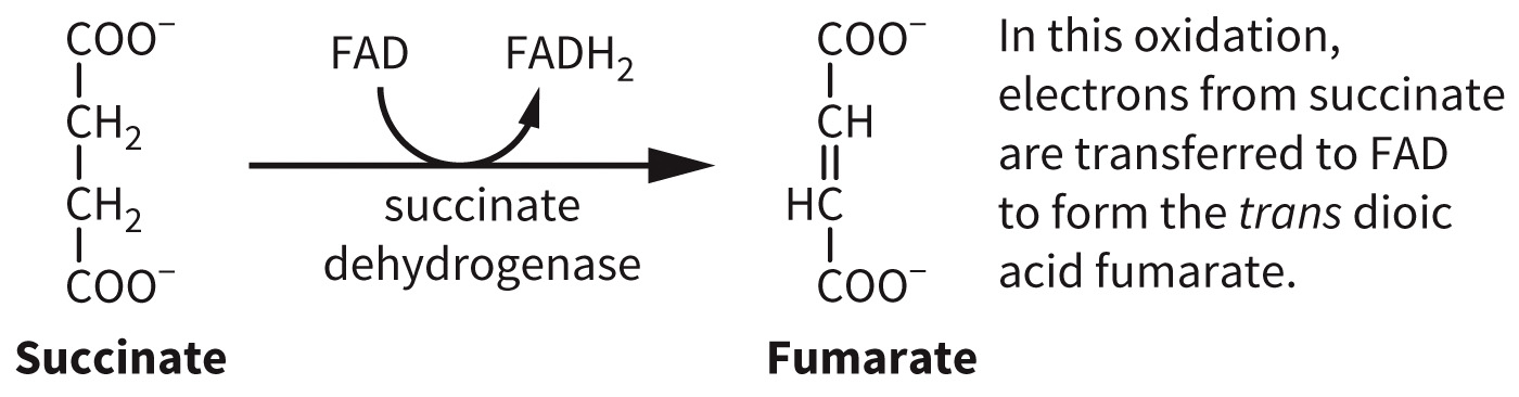
FAD is tightly bound to the enzyme and quickly passes these electrons to ubiquinone to reduced ubiquinone in the electron transport chain.
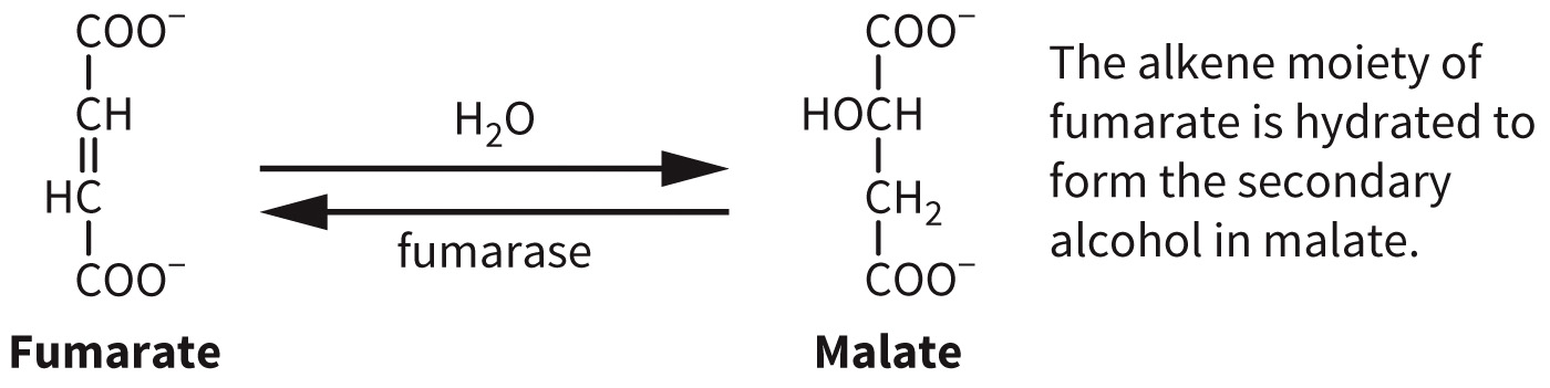
Fumarase
Hydration reaction
Reversible reaction
ATP is neither consumed nor produced.
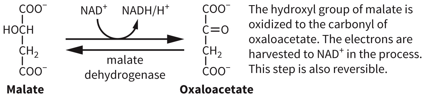
Malate Dehydrogenase
Oxidation reaction
Reversible reaction
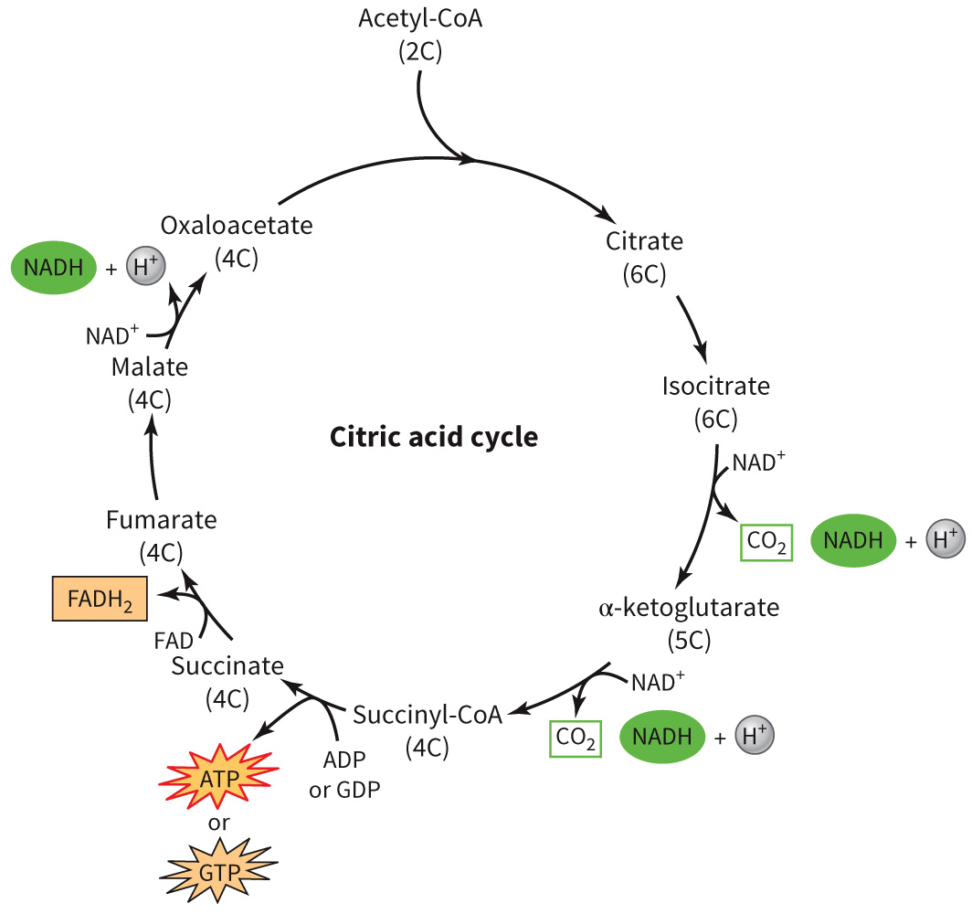
A Schematic View of the Citric Acid Cycle
Figure 7.3 Schematic view of the citric acid cycle.
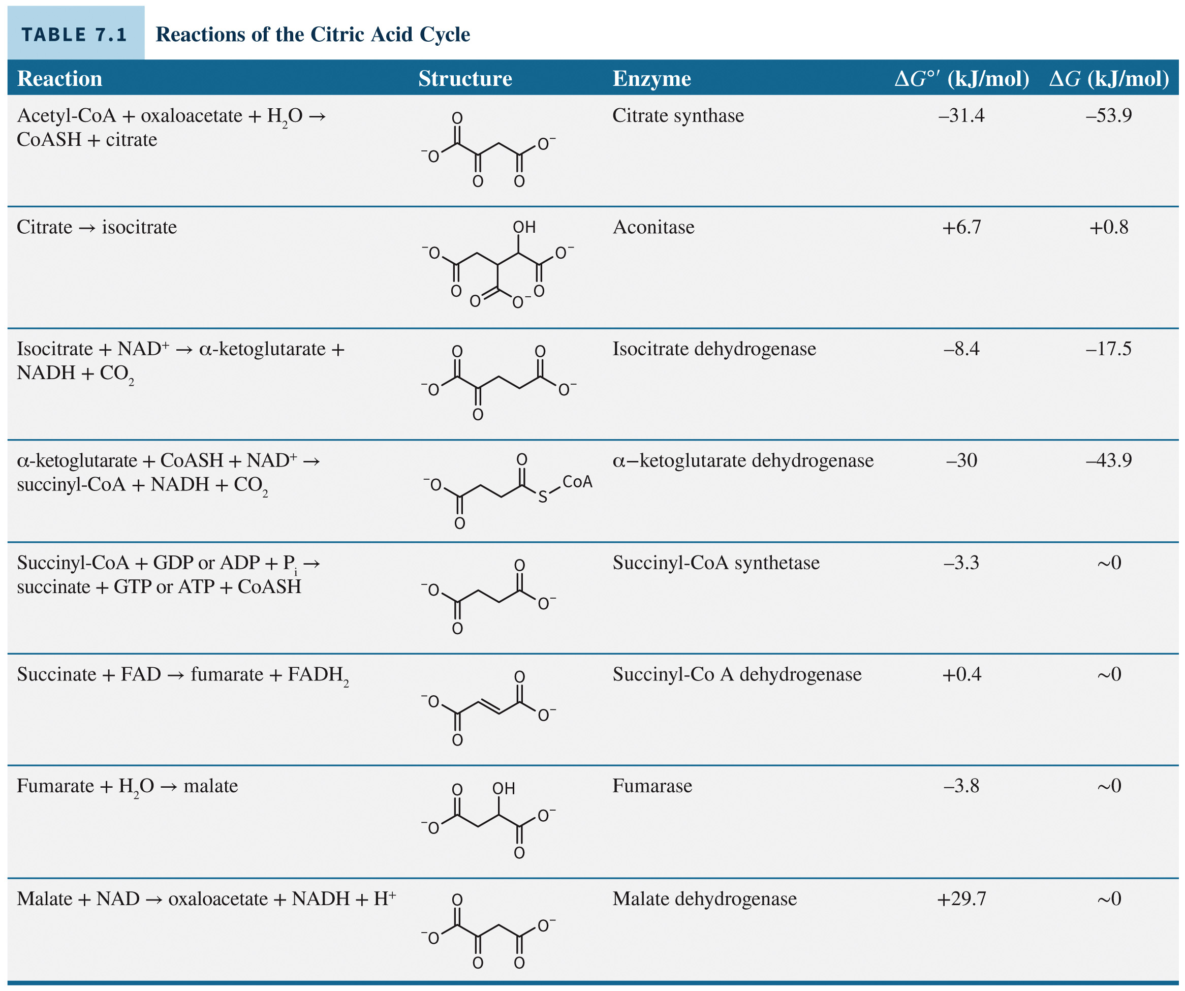
Energetics of the Citric Acid Cycle
Regulation of the Citric Acid Cycle
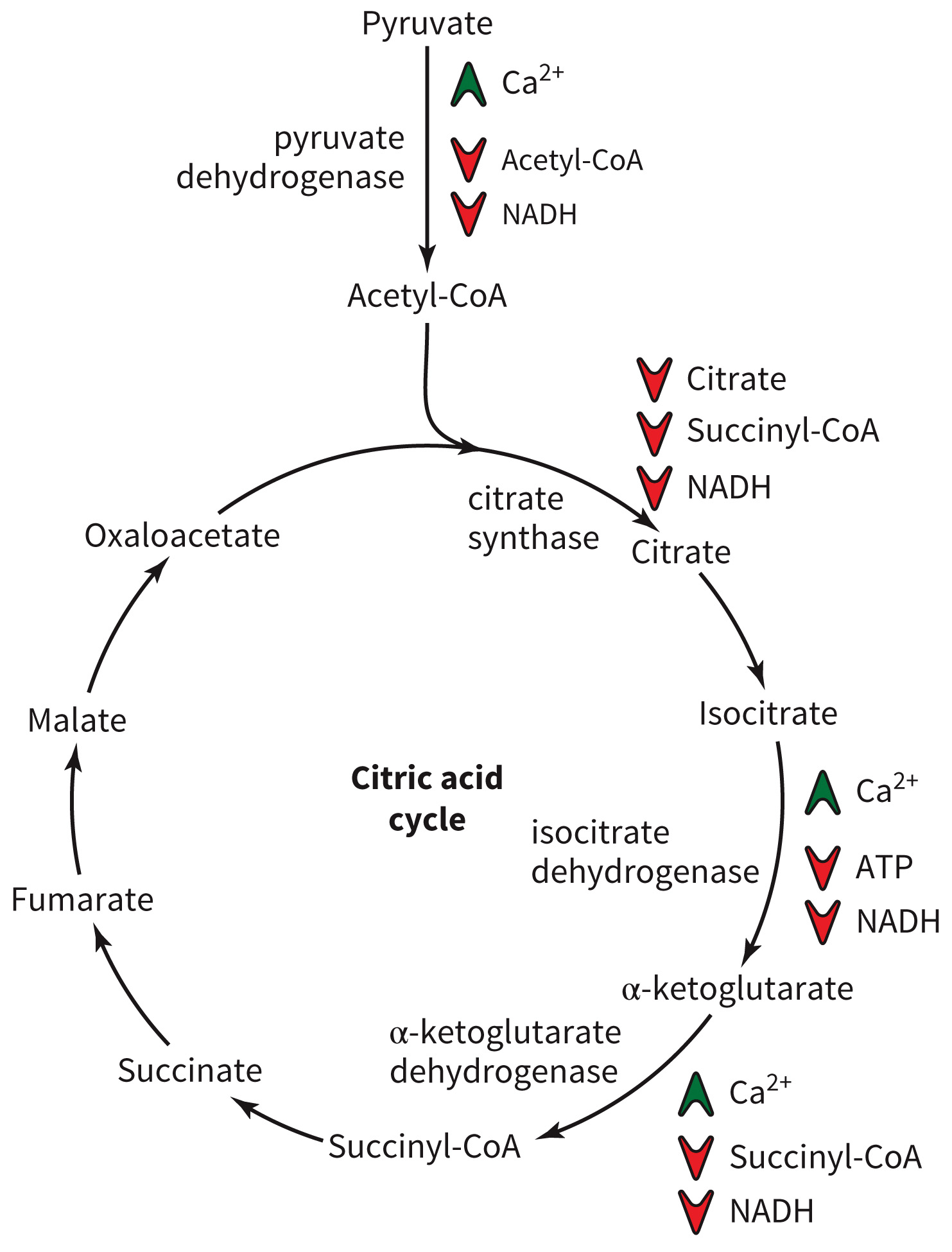
Based on substrate availability
- Increased levels of substrates generally increase flux through the pathway
inhibits three dehydrogenases.
Allosteric regulators: - ATP
- Succinyl CoA
Regulation of the Citric Acid Cycle
Figure 7.4 Regulation of the citric acid cycle.
Anaplerotic Reactions
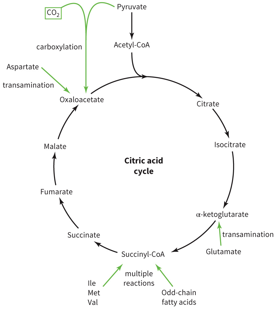
Anaplerotic reactions replenish levels of citric acid cycle intermediates through a number of mechanisms.
Figure 7.5 Central role of the citric acid cycle in metabolism.
Transamination Reactions
Transamination reactions occur when amine groups are shuttled to α-ketoglutarate to generate glutamate and a new α-keto acid from the old amino acid.
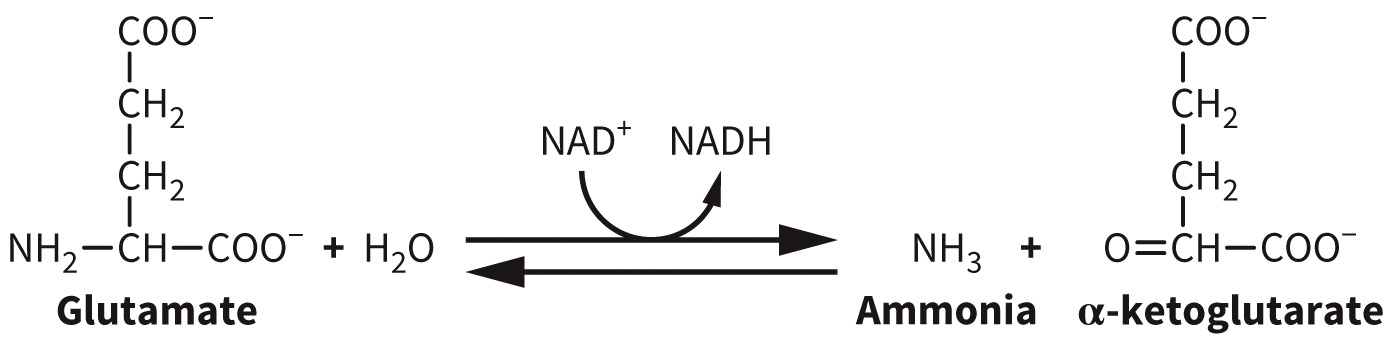
α-Ketoglutarate Replenishment
Levels of α-ketoglutarate can be replenished by removal of the amine moiety from the amino acid glutamate.
Succinyl-CoA Replenishment
Propionyl-CoA can be generated via catabolism of odd-chain fatty acids, branched-chain amino acids (Leu, Ile, Val), or the amino acids Thr and Met (via α-ketobutyrate)
Figure 7.6 Conversion of propionyl-CoA to succinyl-CoA.

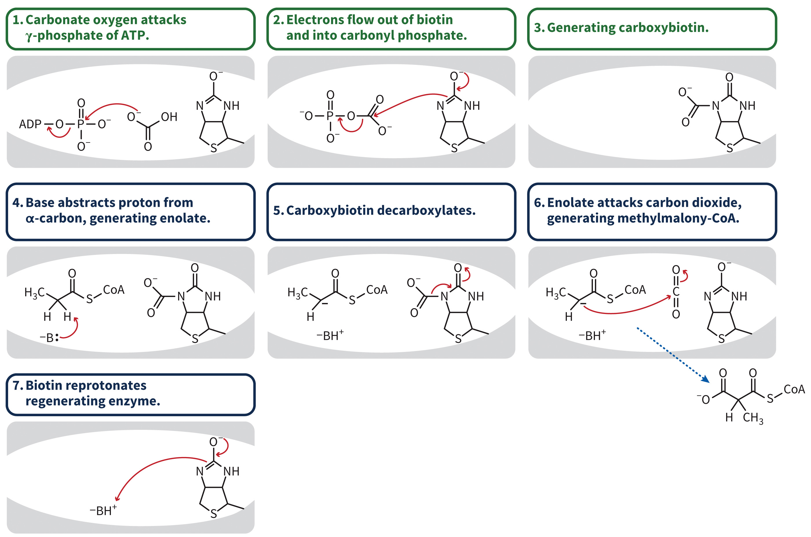
Propionyl CoA Carboxylase Mechanism
Figure 7.7 Mechanism of propionyl-CoA carboxylase.
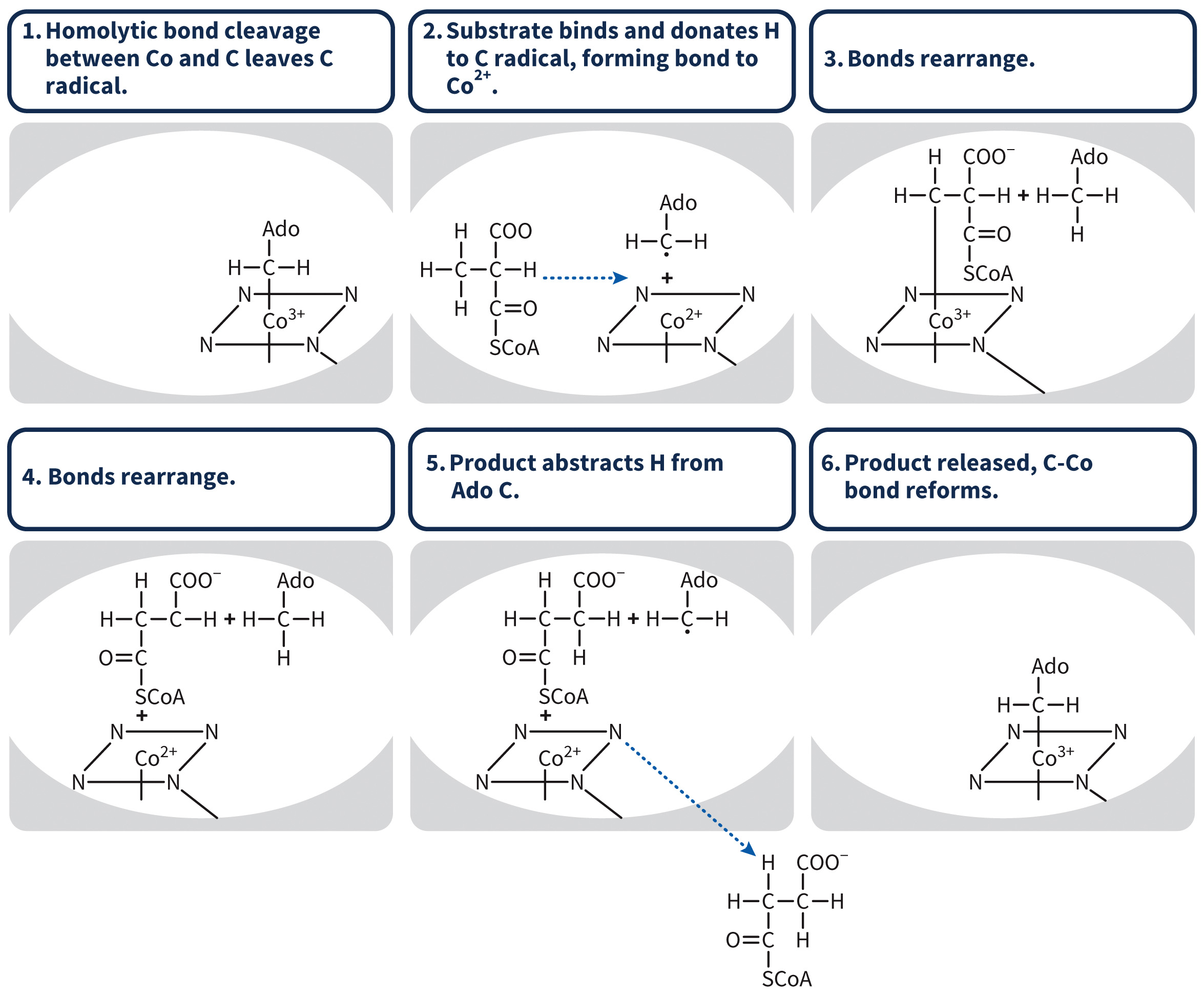
Methyl Malonyl CoA Mechanism
Figure 7.8 Mechansim of methyl malonyl-CoA mutase.
Oxaloacetate Replenishment
Can be produced from pyruvate through gluconeogenesis
Retained in the mitochondrial matrix for use in the TCA cycle
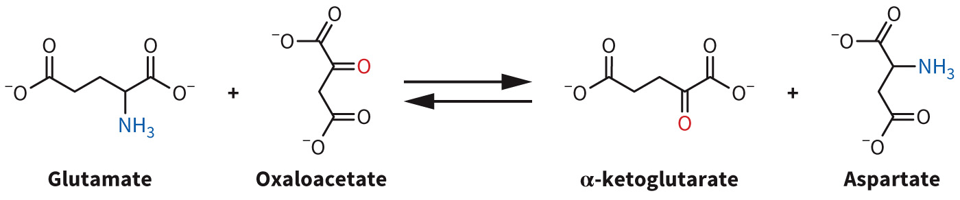
Can be generated from glutamate via aspartate transamination
Pyridoxal phosphate is a cofactor
Figure 7.10 Transamination of aspartate.
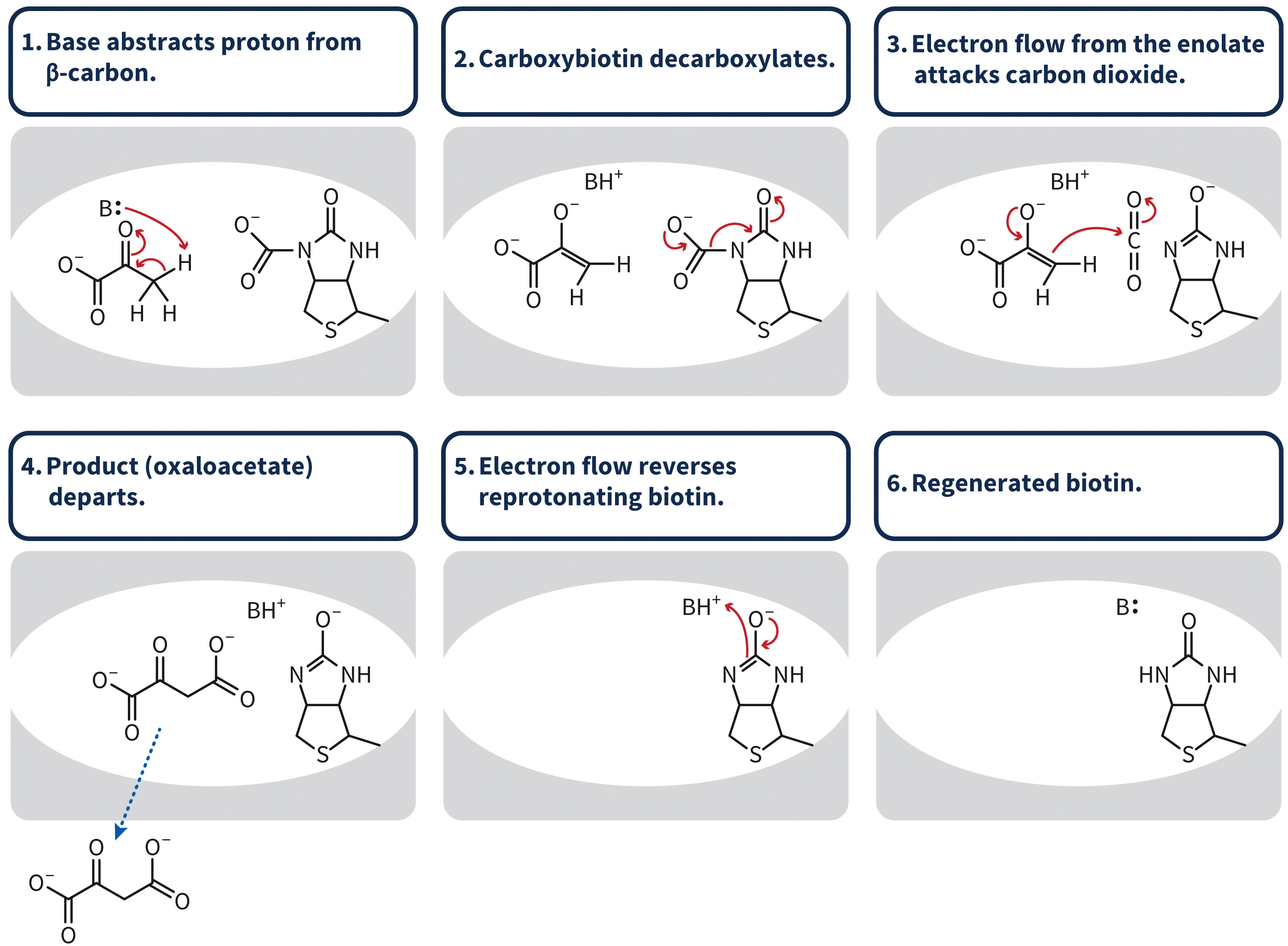
Pyruvate Carboxylase Mechanism
Figure 7.9 Mechanism of pyruvate carboxylase.
Section 7.2 Learning Objective
Illustrate how electron transport results in protons (
Reducing Equivalents
Figure 7.11 Reducing equivalents come from multiple metabolic pathways.
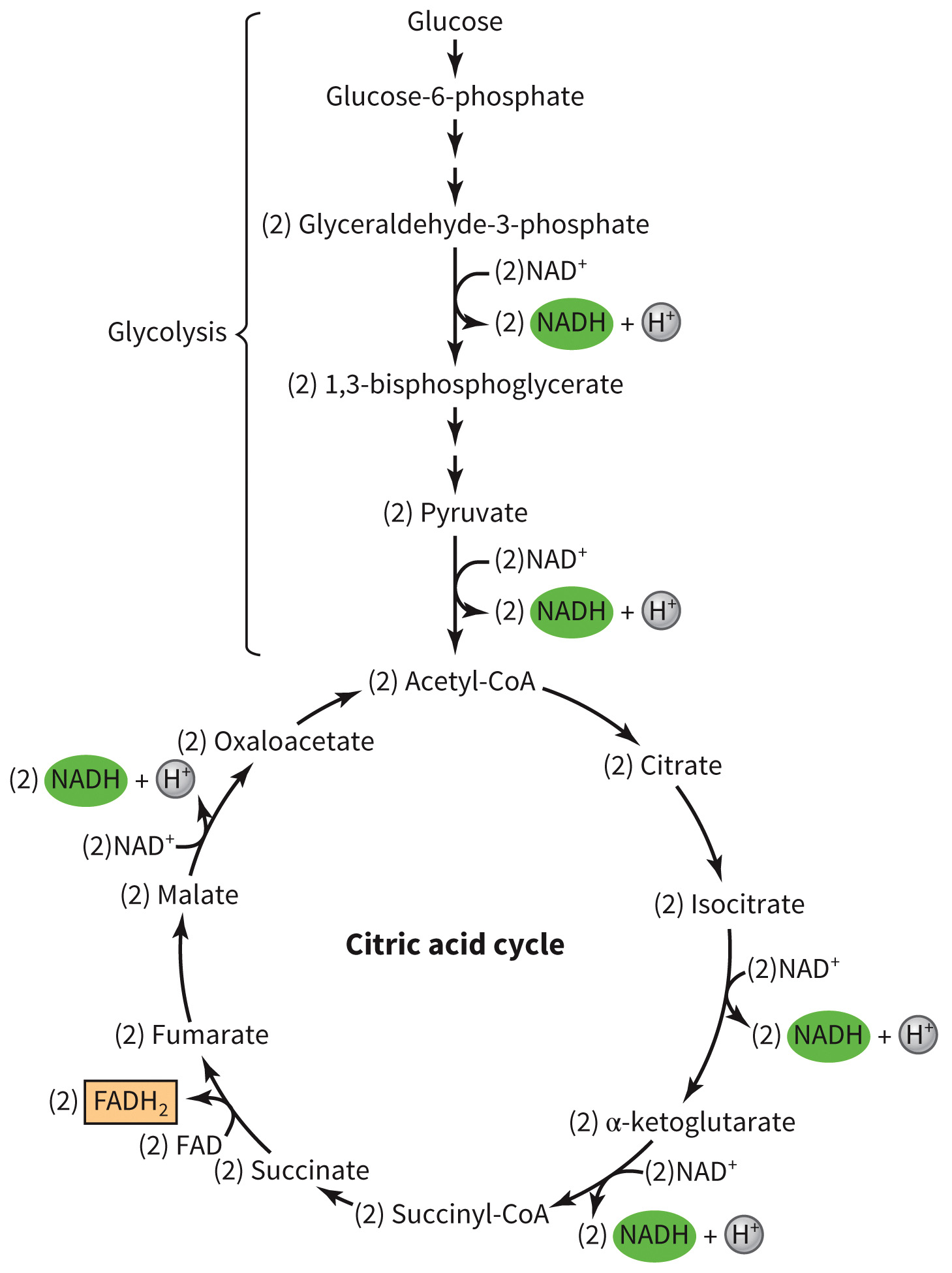
Through these processes – 4 ATP molecules and 10
Electron Transport Chain
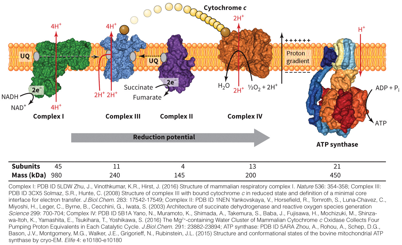
Occurs in the matrix and inner mitochondrial membrane
Figure 7.12 Overview of the electron transport chain.
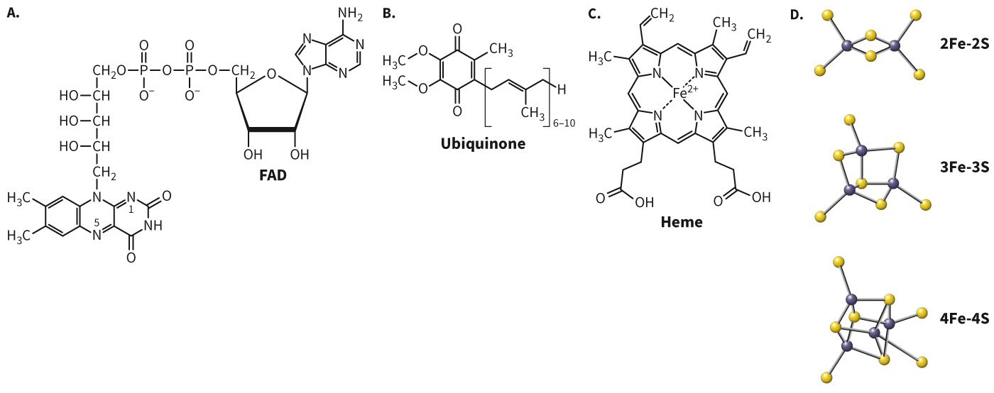
Electron Carriers Involved in the Electron Transport Chain
Figure 7.13 Electron carriers in the electron transport chain.
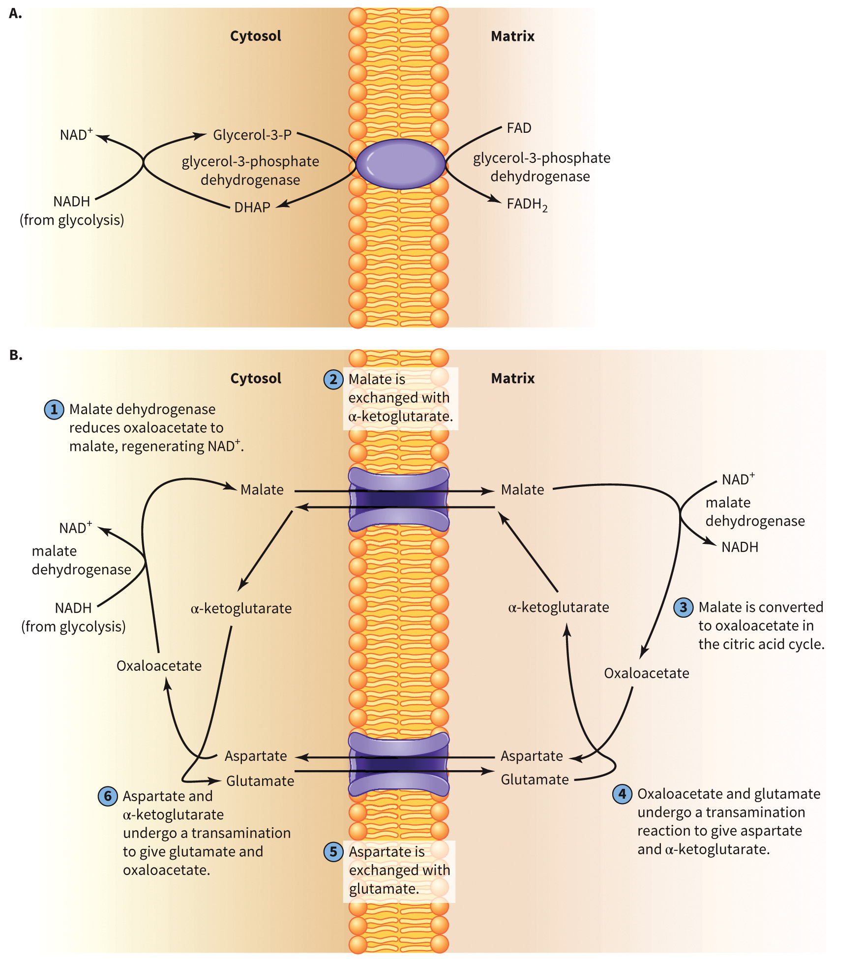
Mitochondrial Shuttles
Two important shuttles that transport metabolites from the mitochondrial matrix to the cytosol
- Glycerophosphate shuttle
- Aspartate-malate shuttle
Mitochondrial Shuttles
Figure 7.14 Mitochondrial shuttles.
Complex I
Site of
Pumps 4
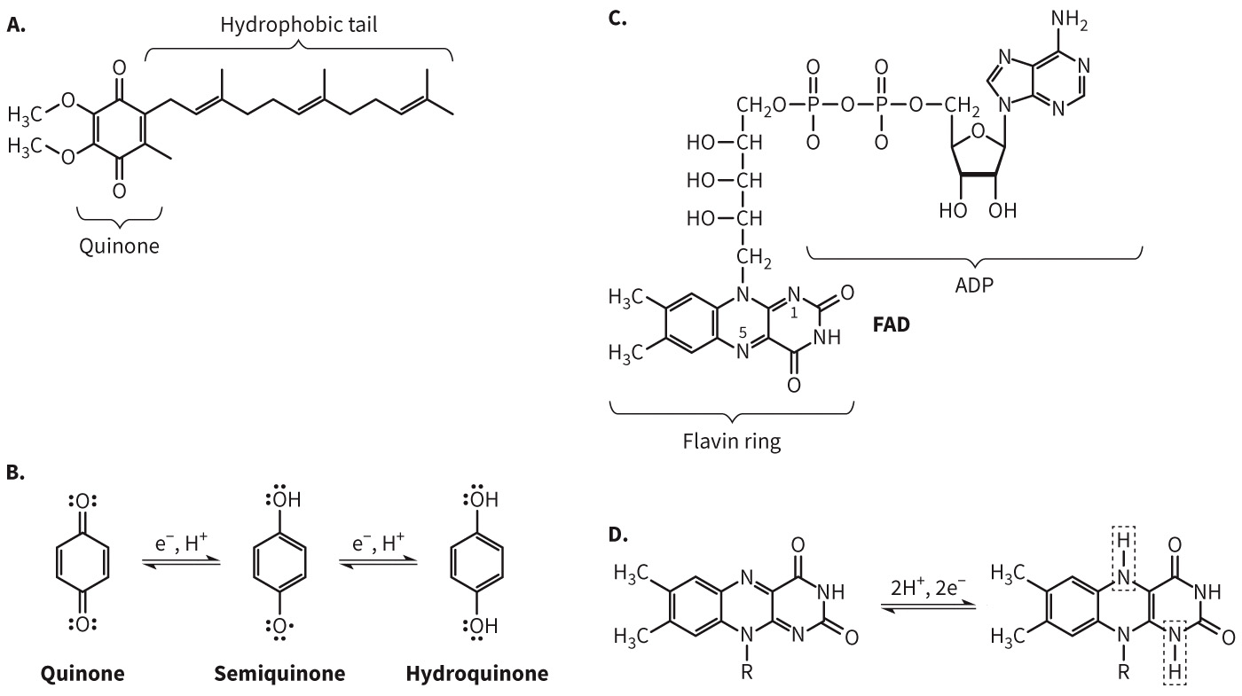
Electrons are transferred to ubiquinone.
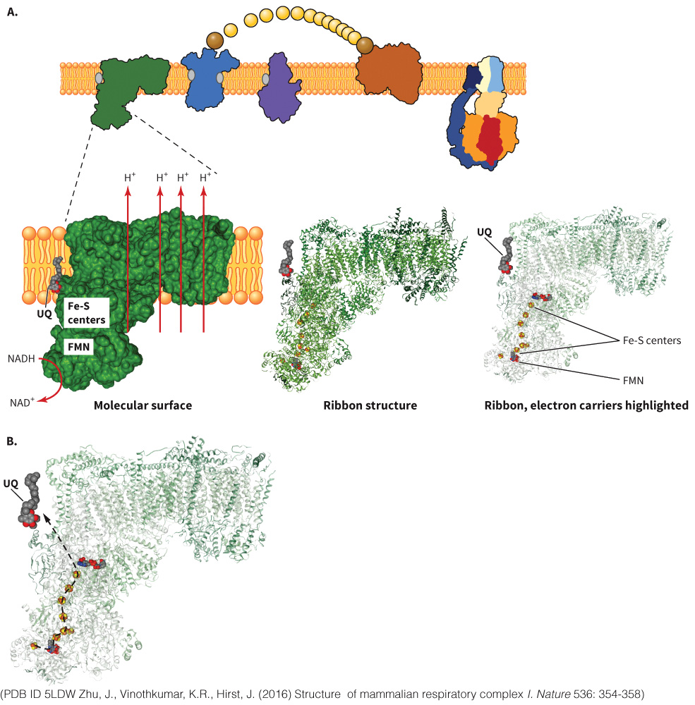
Electron Transport in Complex I
Figure 7.16 Redox states of ubiquinone and FAD.
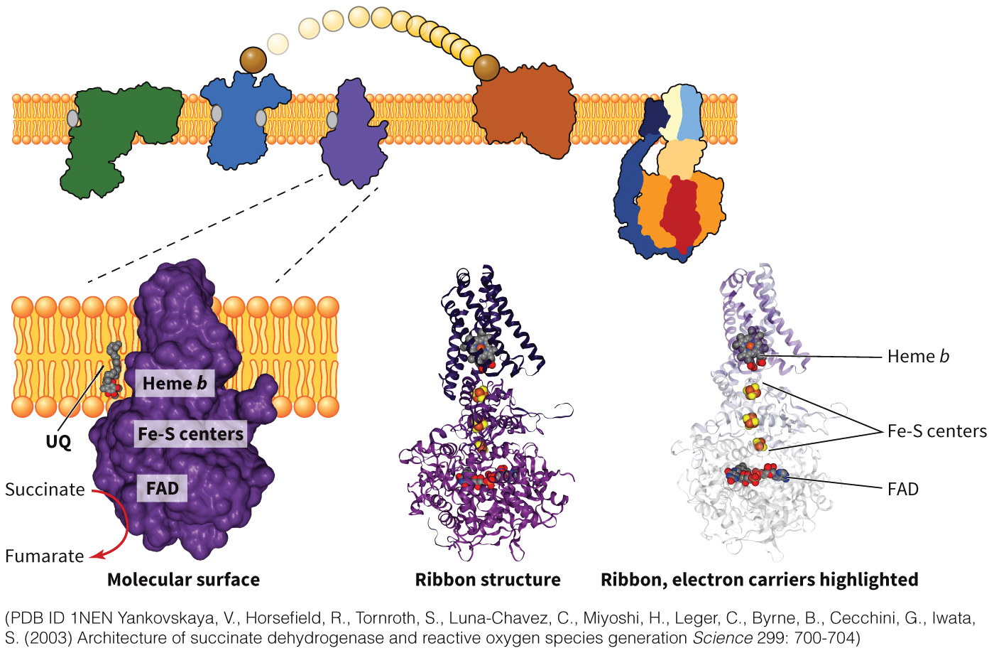
Complex II
Succinate dehydrogenase
Generates
No protons are pumped out of the matrix.
Contains two transmembrane and two hydrophilic domains.
Contains iron-sulfur centers.
Electrons are transferred to Complex III
Figure 7.17A Complex II.
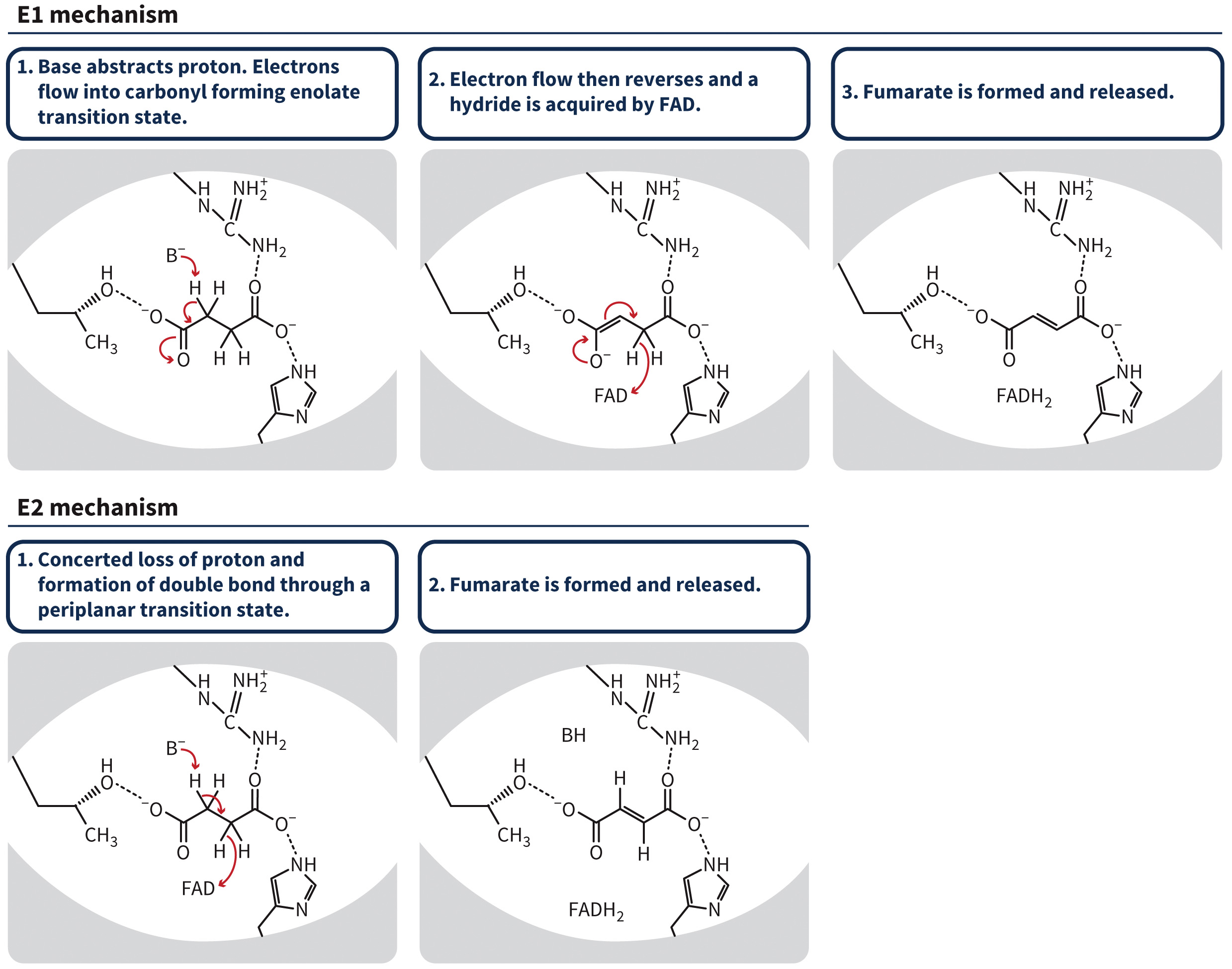
Mechanism for Electron Transport in Complex II
Figure 7.18 Two plausible mechanisms for succinate dehydrogenase.
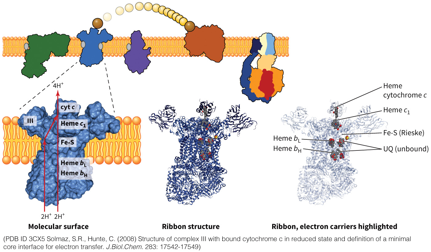
Complex III
Ubiquinone/cytochrome c reductase
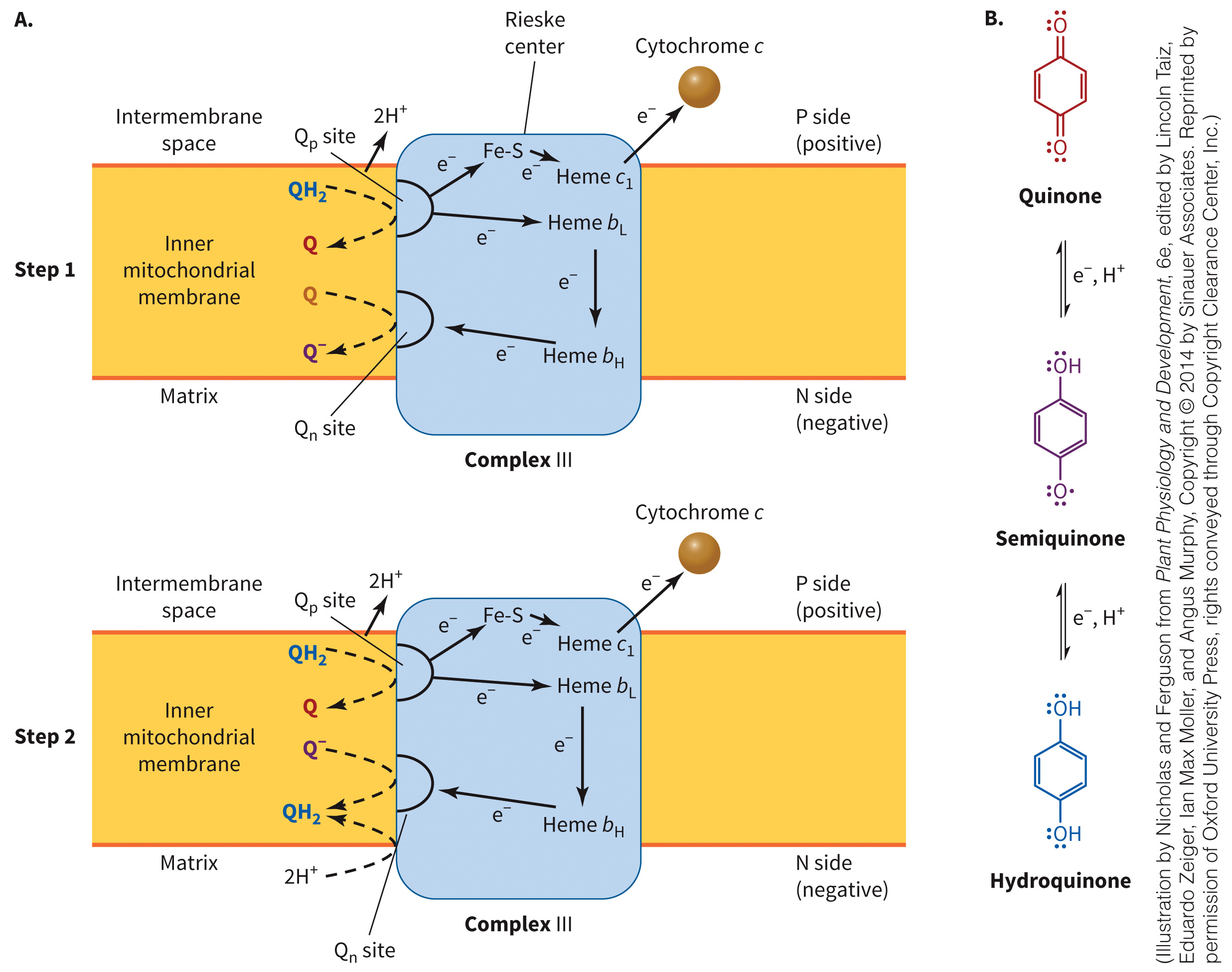
Two ubiquinone binding sites
Heme is involved
Oxidation of one molecule of ubiquinone forms two molecules of reduced cytochrome c
Pumps 4
Figure 7.19A Complex III.
Q Pool Defined
Combination of oxidized and reduced forms of ubiquinone found in the mitochondrial membrane
Q Cycle and Electron Transport
Figure 7.20 Q cycle and electron transport.
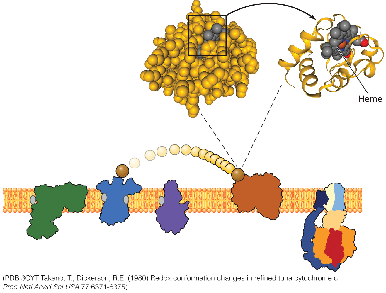
Cytochrome c
A soluble electron carrier
Receives electrons from Complex III
Heme protein that carries one electron in the heme group
Heme is buried in the core of the protein, but is reduced through quantum mechanics
Figure 7.21 Cytochrome c.
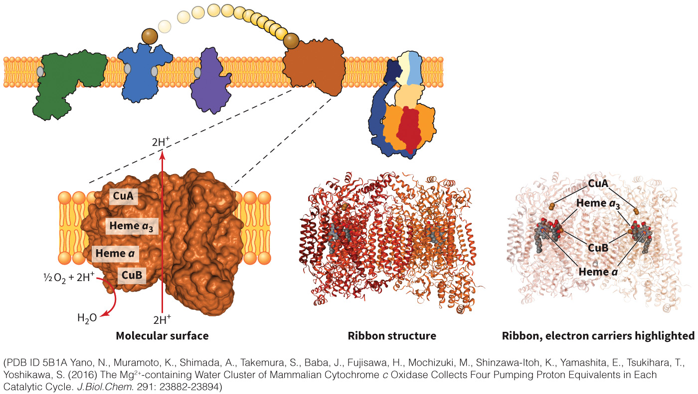
Complex IV
Cytochrome c oxidase
Takes 4
Pumps 2
Reduces
Uses four different electron carriers
Figure 7.22A Complex IV.
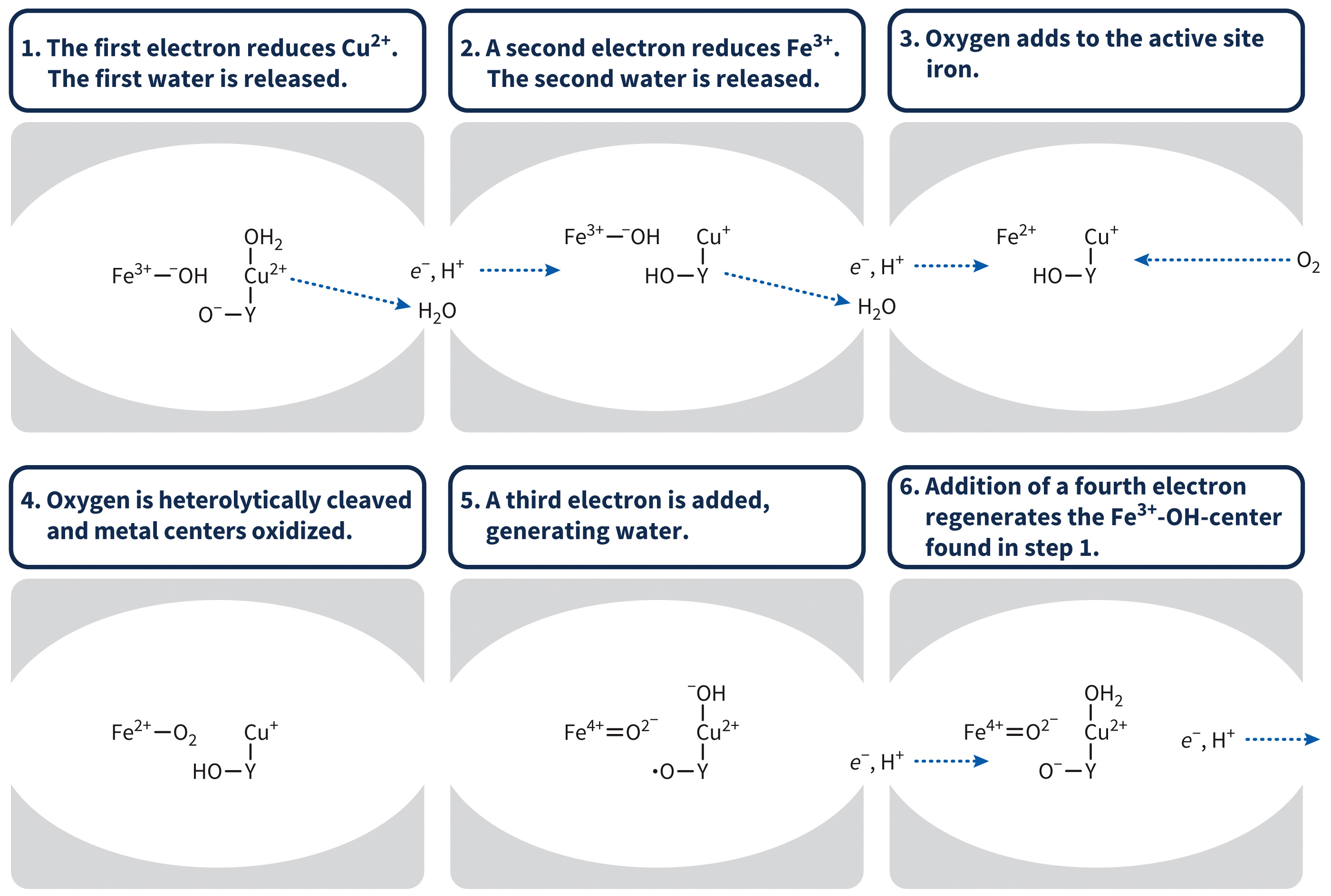
Mechanism for Electron Transport in Complex IV
Figure 7.23 Mechanism of water formation.
Oxygen atom acquires proton from tyrosine, which generates a tyrosyl radical.
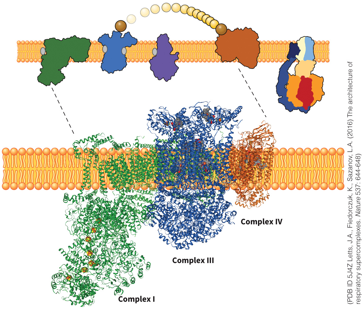
Respirasome Defined
Respirasome is an aggregated supercomplex containing Complexes I, III, and IV.
- Other aggregated complexes include the replisome, transcriptional complex, and signal transduction complexes.
Assists with substrate channeling
Figure 7.24 Respirasome.
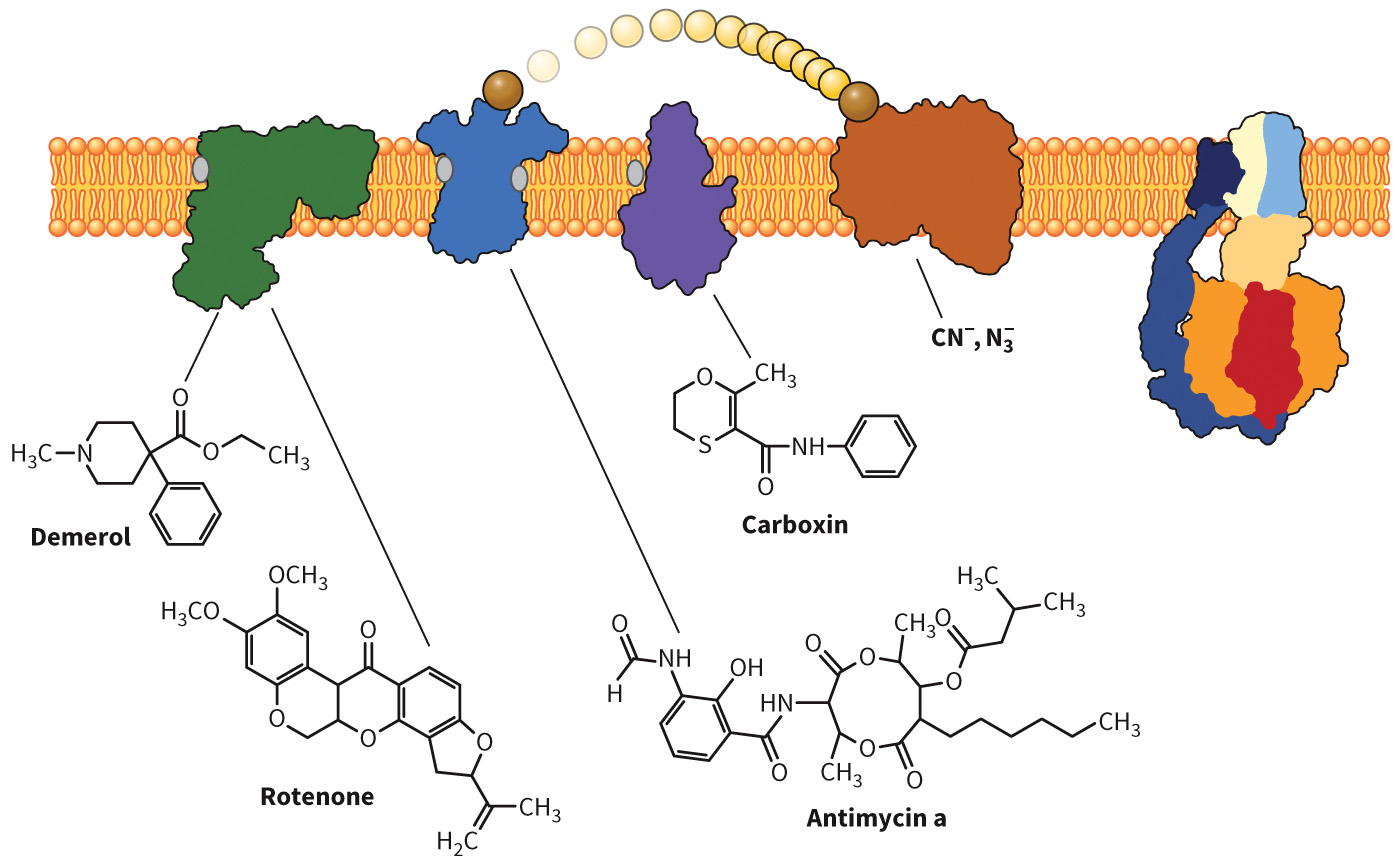
Poisons That Inhibit the Electron Transport Chain
| Drug or poison | Complex | Carrier bound |
|---|---|---|
| Rotenone (insecticide) | I | Fe-S |
| Amytal (amobarbital, barbituates) | ||
| Demerol (meperidine) | ||
| Carboxin (fungicide) | II | Ubiqinone binding site |
| 2-thionyltrifluoroacetone | ||
| Antimycin A1 | III | Cyt bH in the Qn site |
| Cyanide (CN–) | IV | Heme cyt a3 |
| Azide (N3–) | ||
| Carbon monoxide (CO) | ||
| 2,4-dinitrophenol (DNP) | ATP synthase | Uncoupling agent |
| Dicumarol | ||
| FCCP |
Structures of Poisons to the Electron Transport Chain
Figure 7.25 Poisons of electron transport.
Uncouplers and the Electron Transport Chain
Uncouplers interfere with electron transport.
Heat is generated instead of ATP.
DNP is an example of an uncoupler, as is UCP-1 (used by babies and hibernating animals)
Section 7.3 Learning Objective
Describe how the cell uses the electrochemical gradient produced in electron transport to synthesize ATP.
ATP Synthase
Also known as
Contains multiple subunits in both complexes
Enzyme mainly responsible for ATP production
Multimeric enzyme that uses electrochemical energy from the proton gradient to produce ATP from ADP and
Structure of ATP Synthase
Figure 7.26A Reactions of ATP synthase.
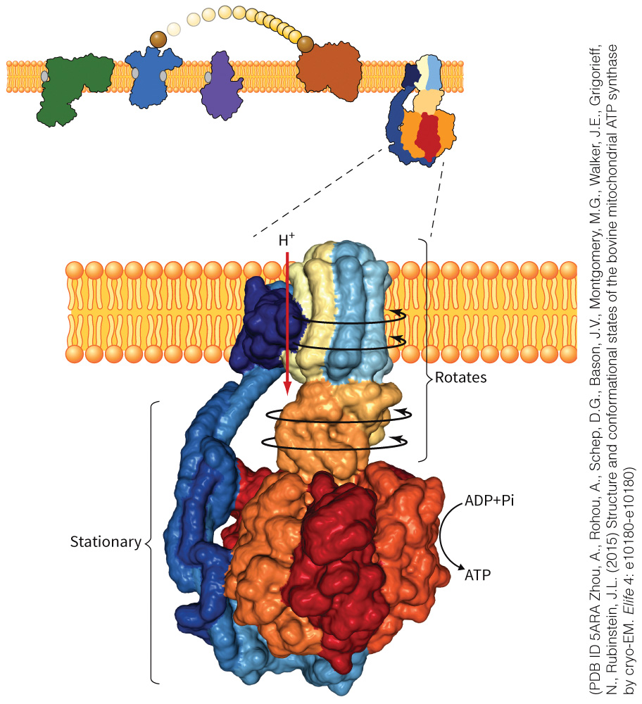
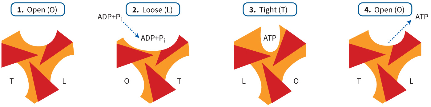
Mechanism of ATP Synthase
Protons move between a and b subunits of
Ring of c subunit rotates.
γ and ε subunits to also move through the hexamer of α and β subunits.
Conformational change occurs.
ATP is formed by binding of ADP and
Open, loose, and tight conformations
ATP Synthase Rotation
Figure 7.26C Reactions of ATP synthase.
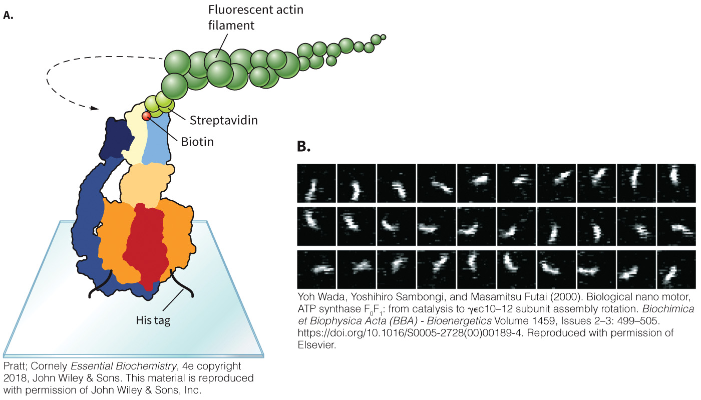
Evidence of ATP Synthase Rotation
Figure 7.27 Rotation of the ATP synthase can be demonstrated in the laboratory.
Proton Motive Force Defined
Proton motive force is the electrochemical potential derived from the uneven distribution of electrons across the inner mitochondrial matrix.
Separated into a chemical and electric potential
ΔG = −2.303 RT ΔpH + nℱ ΔΨm
Chemical Potential Defined
Chemical potential is the higher concentration of protons on the outside of the membrane than on the inside.
Electric Potential Defined
Electric potential is the added positive charge that accumulates on the outside of the membrane.
Measured in V or mV
Voltage drop across the membrane
Gibbs Free Energy Correlation to Membrane Potential
Membrane potential (ΔΨm ) = Ψin
Ψin = voltage inside the membrane
Ψin = voltage outside the membrane
ΔG = nℱΔΨm
ℱ (F, Faraday’s constant) = 96.48 kJ/V · mol
ATP: ADP Translocase Defined
ATP: ADP translocase is an enzyme that transports ATP from the mitochondria in exchange for ADP
Transmembrane domain is an integral membrane protein spanning six α helices
Conformational change occurs
ATPases
Use ATP hydrolysis to perform their proscribed functions in reverse
Five different types
- F-type
- A-type
- V-type
- P-type
- E-type
ATPase Categories
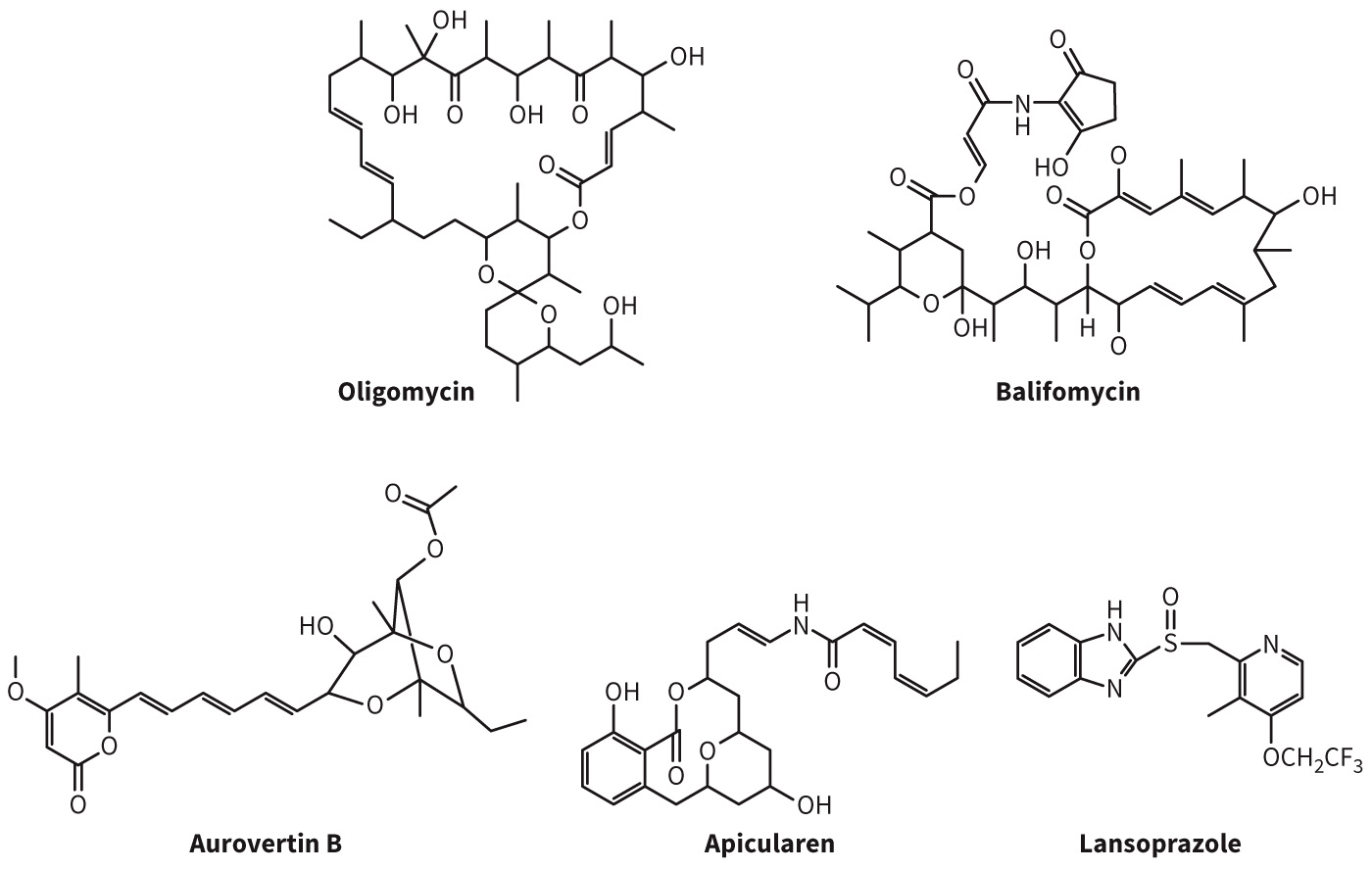
| ATPase | Feature | Inhibitor |
|---|---|---|
| F-type ( | Have isolated factor involved in ADP phosphorylation | Oligomycin Dicyclohexylcarbodiimide (DCC) Aurovertin B |
| A-type | Found in Archea; Similar structure to | |
| V-type | Found in vacuoles; Use energy of ATP hydrolysis to pump | Bafilomycin Concanamycin Apicularen Lobatamide |
| P-type | Pumps that use ATP hydrolysis to move ions from one side of phospholipid bylayer to another; Single transmembrane protein | Omeprazole (Prilosec) Lansoprazole (Prevacid) |
| E-type | Extracellular ATPases; Hydrolyze extracellular ATP and ADP or other nucleotide triphosphates; Involved in platelet aggregation, transplant rejection, and parasite survival |
Structures of Common ATPase Inhibitors